Team:Washington/Gram Negative/Test
From 2010.igem.org
(→SDS-PAGE Protein Array) |
(→Testing the Toxicity of Tse2) |
||
| (61 intermediate revisions not shown) | |||
| Line 25: | Line 25: | ||
</html> | </html> | ||
<!---------------------------------------PAGE CONTENT GOES BELOW THIS----------------------------------------> | <!---------------------------------------PAGE CONTENT GOES BELOW THIS----------------------------------------> | ||
| - | |||
| - | |||
| - | |||
| - | = | + | =Western Blotting Shows Production of Type VI Secretion System Components= |
| - | + | ||
| - | + | ||
| - | + | ||
| - | + | To confirm that our recombineered promoter system resulted in expression of the T6SS in ''E. coli'', we transformed the recombinant fosmid into a T7 expression strain, BL21(DE3), which produces T7 RNA polymerase in the presence of IPTG. We then probed cell extract using a Western blot with antibody against Fha1, an essential component of the T6SS, to verify expressed T6SS protein. The Fha1 protein was expressed under induced conditions, but not under uninduced conditions. This shows that our recombineering was successful and T6SS proteins are being expressed in our new ''E. coli'' system. | |
| - | [[Image:Washington SDS-PAGE.jpg|600px|thumb|center| | + | [[Image:Washington T6SS SDS-PAGE.jpg|600px|thumb|center|'''Western blot for Type VI Secretion Protein. As a control the native fosmid with ''P. aeruginosa'' promoters was expressed in ''P. aeruginosa''. A band showing expressing of T6SS was seen. The engineered fosmid with T7 promoters, K314300, was expressed in ''E. coli''. Expression was induced using 0.5 mM IPTG. A band was seen corresponding to T6SS protein, indicating that the T6SS is being expressed.''']] |
| - | + | ||
| - | = | + | =Western Blotting Shows HSL Induced Expression of Tse2= |
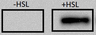
| - | + | In order to determine that Tse2 and Tsi2 were only being produced in the presence of HSL, ''E. coli'' MG1655 containing the [http://partsregistry.org/wiki/index.php?title=Part:BBa_K314202 HSL inducible toxin antitoxin construct] were cultured in liquid LB containing either 10µM HSL, or no HSL. The cultures were pelleted, and then Western blot was performed probing for the presence of Tse2. The cultures grown in HSL+ media showed bands on the Western blot (Figure below) indicative of cells sensing the presence of HSL and producing Tse2. The cultures grown in media without HSL showed no bands, meaning that Tse2 is not being expressed unless HSL is present. This is the expected regulation of our toxin antitoxin circuit. | |
| + | <br> | ||
| + | [[Image:Washington_Tse2_Tsi2_Western.jpg|600px|center|thumb|Western Blot Showing Proper Expression of Tse2]] | ||
| + | |||
| + | =Evidence that Tse2 and Tsi2 are Functioning as a Toxin and Antitoxin System= | ||
| + | With the experiments that we have done so far, it is somewhat difficult to prove beyond a doubt that the Tse2 and Tsi2 proteins are working as expected, since the Tse2 is a toxin and the readout is cell death. We have several points of evidence that imply that Tse2 and Tsi2 are functioning correctly in our hands as a toxin and antitoxin pair. | ||
| + | *Using an IPTG-inducible (non-BioBrick) plasmid we showed that cells with an IPTG-inducible Tse2 failed to grow on plates containing 100 uM IPTG but grew on plates that did not contain IPTG (Data not shown, as this is a replication of previously published work([[#References | [1]]]). This indicates that the Tse2 protein is toxic to cells when expressed. | ||
| + | *Although we obtained a [http://partsregistry.org/Part:BBa_K314200 promoter-free Tse2 BioBrick], we were never able to clone Tse2 alone downstream of the F2620 promoter without an inactivating mutation somewhere in the F2620 or Tse2 gene. This implies that even at the low, leaky level of expression of the F2620, Tse2 is toxic to cells. | ||
| + | *When paired with the antitoxin Tsi2, we were readily able to clone Tse2 downstream of F2620 with no mutations, and expression of protein confirmed with anti-Tse2 antibody. Cell survival in this case is evidence that the Tsi2 antitoxin is working in our system. | ||
| + | |||
| + | =Future Work and Characterization= | ||
| + | |||
| + | ==Testing the Complete Theraputic System== | ||
| + | We were able to show that the T6SS is being expressed in ''E. coli'', and that Tse2(the toxin) is expressed only when HSL is present. To show that the entire system works will require a competition assay in which our engineered ''E. coli'' probiotic, expressing the T6SS, kills target Gram-negative cells by injecting Tse2 but only in the presence of HSL. We would expect our probiotic to outcompete wild type ''E. coli'' when both the T6SS and Tse2 Tsi2 operon are being expressed (+HSL,+IPTG), but not when only the T6SS ( -HSL,+IPTG) or only Tse2 and Tsi2 (+HSL, -IPTG) are being expressed. | ||
| + | |||
| + | ==Testing the Toxicity of Tse2== | ||
| + | We intend to determine the relative level of Tse2 expression required to cause cell death, and to compare the toxicity of [http://partsregistry.org/wiki/index.php?title=Part:BBa_K314200 Tse2] to [http://partsregistry.org/Part:BBa_P1010 ccdB] (the most commonly used cell death protein). This would require that both Tse2 and CcdB be placed downstream from the same inducible promoter. Cell growth curves would then be measured at various levels of the inducer. The toxin that is more toxic would kill at a lower concentration of the inducer molecule (and thus lower protein levels). We had intended to place both Tse2 and CcdB downstream from [http://partsregistry.org/Part:BBa_F2620 F2620] and compare growth at various HSL concentrations, but we were unable to obtain sequence verified F2620.Tse2 constructs due to mutations. This implies that the leakiness from the pLux promoter is enough to create a selection advantage against even the smallest levels of Tse2 production. Conducting this test would require the use of a less leaky promoter (such as an IPTG-inducible promoter) that would only produce Tse2 when the inducer is present. | ||
| + | |||
| + | ==References== | ||
| + | 1. Hood RD, Singh P, Hsu F, Güvener T, Carl MA, Trinidad RR, Silverman JM, Ohlson BB, Hicks KG, Plemel RL, Li M, Schwarz S, Wang WY, Merz AJ, Goodlett DR, Mougous JD. [http://www.cell.com/cell-host-microbe/abstract/S1931-3128%2809%2900417-X#Summary A type VI secretion system of Pseudomonas aeruginosa targets a toxin to bacteria]. Cell Host Microbe. 2010 Jan 21;7(1):25-37. PMID: 20114026 | ||
<!---------------------------------------PAGE CONTENT GOES ABOVE THIS----------------------------------------> | <!---------------------------------------PAGE CONTENT GOES ABOVE THIS----------------------------------------> | ||
<div style="text-align:center"> | <div style="text-align:center"> | ||
| - | '''← [[Team:Washington/ | + | '''← [[Team:Washington/Gram Negative/Build|Building the Gram(-) Therapeutic]]''' |
| | ||
| | ||
| | ||
| - | '''[[Team:Washington/ | + | '''[[Team:Washington/Gram Positive|Overview of Gram(+) Therapeutic]] →''' |
</div> | </div> | ||
{{Template:Team:Washington/Templates/Footer}} | {{Template:Team:Washington/Templates/Footer}} | ||
Latest revision as of 16:50, 26 October 2010
Western Blotting Shows Production of Type VI Secretion System Components
To confirm that our recombineered promoter system resulted in expression of the T6SS in E. coli, we transformed the recombinant fosmid into a T7 expression strain, BL21(DE3), which produces T7 RNA polymerase in the presence of IPTG. We then probed cell extract using a Western blot with antibody against Fha1, an essential component of the T6SS, to verify expressed T6SS protein. The Fha1 protein was expressed under induced conditions, but not under uninduced conditions. This shows that our recombineering was successful and T6SS proteins are being expressed in our new E. coli system.

Western Blotting Shows HSL Induced Expression of Tse2
In order to determine that Tse2 and Tsi2 were only being produced in the presence of HSL, E. coli MG1655 containing the [http://partsregistry.org/wiki/index.php?title=Part:BBa_K314202 HSL inducible toxin antitoxin construct] were cultured in liquid LB containing either 10µM HSL, or no HSL. The cultures were pelleted, and then Western blot was performed probing for the presence of Tse2. The cultures grown in HSL+ media showed bands on the Western blot (Figure below) indicative of cells sensing the presence of HSL and producing Tse2. The cultures grown in media without HSL showed no bands, meaning that Tse2 is not being expressed unless HSL is present. This is the expected regulation of our toxin antitoxin circuit.
Evidence that Tse2 and Tsi2 are Functioning as a Toxin and Antitoxin System
With the experiments that we have done so far, it is somewhat difficult to prove beyond a doubt that the Tse2 and Tsi2 proteins are working as expected, since the Tse2 is a toxin and the readout is cell death. We have several points of evidence that imply that Tse2 and Tsi2 are functioning correctly in our hands as a toxin and antitoxin pair.
- Using an IPTG-inducible (non-BioBrick) plasmid we showed that cells with an IPTG-inducible Tse2 failed to grow on plates containing 100 uM IPTG but grew on plates that did not contain IPTG (Data not shown, as this is a replication of previously published work( [1]). This indicates that the Tse2 protein is toxic to cells when expressed.
- Although we obtained a [http://partsregistry.org/Part:BBa_K314200 promoter-free Tse2 BioBrick], we were never able to clone Tse2 alone downstream of the F2620 promoter without an inactivating mutation somewhere in the F2620 or Tse2 gene. This implies that even at the low, leaky level of expression of the F2620, Tse2 is toxic to cells.
- When paired with the antitoxin Tsi2, we were readily able to clone Tse2 downstream of F2620 with no mutations, and expression of protein confirmed with anti-Tse2 antibody. Cell survival in this case is evidence that the Tsi2 antitoxin is working in our system.
Future Work and Characterization
Testing the Complete Theraputic System
We were able to show that the T6SS is being expressed in E. coli, and that Tse2(the toxin) is expressed only when HSL is present. To show that the entire system works will require a competition assay in which our engineered E. coli probiotic, expressing the T6SS, kills target Gram-negative cells by injecting Tse2 but only in the presence of HSL. We would expect our probiotic to outcompete wild type E. coli when both the T6SS and Tse2 Tsi2 operon are being expressed (+HSL,+IPTG), but not when only the T6SS ( -HSL,+IPTG) or only Tse2 and Tsi2 (+HSL, -IPTG) are being expressed.
Testing the Toxicity of Tse2
We intend to determine the relative level of Tse2 expression required to cause cell death, and to compare the toxicity of [http://partsregistry.org/wiki/index.php?title=Part:BBa_K314200 Tse2] to [http://partsregistry.org/Part:BBa_P1010 ccdB] (the most commonly used cell death protein). This would require that both Tse2 and CcdB be placed downstream from the same inducible promoter. Cell growth curves would then be measured at various levels of the inducer. The toxin that is more toxic would kill at a lower concentration of the inducer molecule (and thus lower protein levels). We had intended to place both Tse2 and CcdB downstream from [http://partsregistry.org/Part:BBa_F2620 F2620] and compare growth at various HSL concentrations, but we were unable to obtain sequence verified F2620.Tse2 constructs due to mutations. This implies that the leakiness from the pLux promoter is enough to create a selection advantage against even the smallest levels of Tse2 production. Conducting this test would require the use of a less leaky promoter (such as an IPTG-inducible promoter) that would only produce Tse2 when the inducer is present.
References
1. Hood RD, Singh P, Hsu F, Güvener T, Carl MA, Trinidad RR, Silverman JM, Ohlson BB, Hicks KG, Plemel RL, Li M, Schwarz S, Wang WY, Merz AJ, Goodlett DR, Mougous JD. [http://www.cell.com/cell-host-microbe/abstract/S1931-3128%2809%2900417-X#Summary A type VI secretion system of Pseudomonas aeruginosa targets a toxin to bacteria]. Cell Host Microbe. 2010 Jan 21;7(1):25-37. PMID: 20114026
 "
"

