ApoControl Notebook
What we did
Short description of our work, our results and our supporters
The creation of certain constructs was necessary for our two systems for cell selection by means of apoptosis: “Cut’N’Survive” and “Jump-Or-Die”. We searched for sources of the DNA sequences we needed and found several supporters which are listed below.
Most genes and promoters were amplificated via PCR with overhang-primers with the BioBrick prefix or suffix. If the sequence contained a EcoR1-, Pst1-, Xba1-, Spe1- or Not1- restriction site, we used mutagenesis primers and fusioned both DNA parts by fusion PCR. All PCRs worked out, even the fusion PCRs.
The length of the PCR products were tested by agarose gel electrophoresis. We tried to sequence our PCR products, but obtained poor results and resorted to sequencing the plasmids.
In parallel, we made competent cells and multiplied ccdB (death gene)-vectors with different antibiotic resistances. All components were digested with the appropriate restriction enzymes. The samples were cleaned with a PCR clean up kit or dephosphorylated to reduce false ligations.
We ligated our constructs and several interim stages with the 3A-assembly according to our schedule. The ligations were transformed to E.coli DH5α strains and selected by antibiotics. Afterwards, some colonies were picked and we tested the insertion of the construct by colony PCR.
If the colony PCR resulted in bands of the right size, we extracted the plasmids from overnight cultures and sequenced the samples with forward and reverse BioBrick primers.
Unfortunately, not all BioBricks were cloned succesfully. However, we were able to produce 4 BioBricks, one of which represents a full construct while the other three are intermediates. The system wasn't completed on time, so we weren´t able to test them in eukarytic cell lines.
The protocols we used are listed here: Protocols
These Biobricks we submitted:
- BBa_K368004: attP+eGFP+SV40PA
- BBa_K368011: eGFP+SV40PA
- BBa_K368016: TEVrecognition site+N-degron+SF3b155
- BBa_K368019: TEV-Protease+p14*+TEVrecognition site
Sources, helpers and supporters:
- Prof. Dr. Angelika Böttger :
- prevTRE (tet-on CMV promoter; inducible by doxycycline in special cell lines)
- supported the construction ideas and would have given us the cells and mediums we would have needed
- SV40PA (Polyadenylation site): gave us a vector containing it
- Human Bak: her assistant Erika Clement gave us appropriate cDNA
Contents>
8-10-2010
Transforming competent cells
- eGFP Biobrick: BBa_I714891 SDY_eGFP (Kanamycin)
- TEV recogn N Degron SF3 = pDS7 (Ampicillin)
- TEV p14 recogn = 190-6 (Ampicillin)
-> Protocol: (3 Transformation)
- We added 2 µl DNA
- We plated out 200 µl
Plasmid Isolation
- CMV-Promoter Biobrick: BBa_J52034
-> Protocol:(4 Plasmid extraction from cells)
- Prepared overnight culture, measured concentration of DNA
-> Poor results -> thrown away
8-11-2010
New Plasmid Extraction
- CMV-Promoter Biobrick: BBa_J52034
-> Protocol: (4 Plasmid extraction from cells)
- Plasmid concentration: 143ng/µl
Prepared overnight culture of eGFP BBa_I714891
- 3 ml LB-Media + 4 µl Kanamycin
- Inoculated with 1 colony of BBa_I714891 -> 37°C
Prepared overnight culture of 190-6 and pDS7 and eGFP (BBa_I714891) in falcons
- for 190-6 and pDS7: 10µl Ampicillin + 10 ml LB-Media + colony of plate
- for eGFP: 13,3 µl Kanamycin + 10 ml LB-Media + 1 colony of plate
Restriction digestion of CMV-Promoter BBa_J52034 with EcoRI and PstI
| H2Oddest, sterile
| 10,3 µl
|
| RE10 + Buffer H
| 2,0 µl
|
| acetylated BSA (18ng/µl)
| 0,2 µl
|
| DNA (0,143µg/µl)
| 6,0 µl
|
-> mixed
- plus: EcoRI (10µg/µl): 0,5 µl resp. PstI (10µg/µl): 0,5 µl
- incubated at room temperature from 12:10 to 15:00, 1 hour at 37°C, 2 hours at 60°C
- frozen at -20°C
Prepare new/fresh overnight culture of CMV-Promoter Biobrick: BBa_J52034
- 1 ml of "old" culture + 3 ml LB-Media + 4 µl Kanamycin -> 37°C
8-12-2010
Plasmid Extraction of pDS7, eGFP, 190-6
-> Protocol: (4 Plasmid extraktion from cells)
- pDS7 (458ng/µl), eGFP (55ng/µl), 190-6 (193ng/µl)
Restriction digest of pDS7, eGFP, 190-6
- with EcoRI and PstI in buffer H (for testing DNA is correct)
-> Protocol: (5 Restriction digest)
- 10µg DNA: pDS7 (2µl), eGFP (15µl), 190-6 (10µl)
Plate colonies for plasmid extraction
- CMV (Kanamycin), eGFP (Kanamycin), pDS7 (Ampicillin), 190-6 (Ampicillin))
- PhiC31o plated on Ampicillin-Agar, stored at 37°C
50% Glycerol made
- for PhiC31o glycerol stock (produced later)
8-13-2010
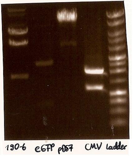 Gelfoto from the EcoR1 and Pst1 Restrictiondigest of 190-6, eGFP, pDS7 and CMV Inoculate CMV into LB medium with ampicillin
- CMV (BBa_J52034) from 10.8.2010 inoculated into LB medium with ampicillin, as falsly inoculated in Kanamycin
Agarosegelelectrophoresis with digestions
->Protocol (11 Agarose gel electrophoresis)
- Agarosegelelectrophoresis with the digestions (CMV, eGFP, pDS7, 190-6), 125V for 30 minutes and then for 20 minutes;
- expected DNA bands: 190-6 (4840bp, 1903bp), pDS7 (8027bp, 6bp), CMV (654 bp (Insert), 2079bp (Plasmid)), eGFP (720bp (Insert), 2750bp (Plasmid))
- Correct DNA bands for 190-6 (~4800bp, ~1900bp, ~6700bp (undigested plasmid)) and eGFP (~2000bp (Plasmid), ~750 bp (Insert)); CMV probably not digested (two bands; one probably normal, one supercoiled) and pDS7 not clear
Restriction digest from CMV and pDS7
-> Protocol (5 Restriction digest)
- Restriction digest from CMV (EcoR1, Pst1; 6µl DNA, buffer H) and pDS7 (EcoR1, Spe1; 2µl DNA, buffer B)
Agarosegelectrophoresis with digestions
->Protocol (11 Agarose gel electrophoresis)
- Agarosegelelectorphoresis for 30 minutes, 150V
- Expected DNA bands: CMV see above, pDS7 (3647bp, 3369bp, 1011bp, 6bp)
- false DNA bands CMV (~1200 bp, ~2000 bp) and pDS7 (~8000bp two bands, ~1100 bp); required to isolate a new colony for these two Plasmidextractions
Plated CMV on Ampicllin-Agar
- Plated the colony from CMV (BBa_J52034) for Plasmidextraction (Ampicillin), as falsly plated on Kanamycin
8-14-2010
weekend
8-15-2010
weekend
8-16-2010
Planting colonies
- transfer 1 ml PhiC31o culture to new LB medium + Amp, 37°C
- pick up CMV and pDS7 colonies from plates and transfer to LB medium+Amp, 37°C
Plasmid Extraction of PhiC31o
->Protocol (4 Plasmid extraktion from cells)
- plasmid extraction of PhiC310
->27,5ng/µl DNA and second plasmid extraction of PhiC310 (i. o. to get more DNA); first eluation-step with first eluation-extraction
-> 60ng/µl DNA
Restriction digest
->Protocol (5 Restriction digest)
- restriction digest of PhiC310 with EcoR1 and Spe1
| H2Oddest, sterile
| 0 µl
|
| Buffer B
| 2,0 µl
|
| BSA (1:10)
| 2 µl
|
| DNA (0,06µg/µl)
| 15,0 µl
|
| EcoR1
| 0,5 µl
|
| Spe1
| 0,5µl
|
restriction digest in the thermo cycler (program "Verdau", see protocol)
Handling primers after arrival (1,2,3,4,5,6,11,12)
->Protocol (9 Handling primers)
PCR preparations
- 10mM dNTP mix made from 100 mM dATP, dGTP, dCTP, dTTP by taking 100µl of each and adding 600µl H 2 O
PCR 1 and 6
- PCR of the tet inducible CMV minimal promotor out of prevTRE (=PCR 1 with Primer 1 and 2) and SV40PA out of pcDNA3 (=PCR 6 with Primer 11 and 12)
->Protocol (10 PCR with Pfu)
Mixture:
|
| pTRERev (0,15µg/µl)
| pcDNA3 (0,6 µg/µl)
|
| Primer
| 2*2,5µl (P1+P2)
| " (P11+P12)
|
| 300ng template
| 0,5µl
| 2µl
|
| 10x Buffer Pfu
| 5µl
| "
|
| dNTP Mix
| 1µl
| "
|
| Pfu Polymerase (3u/µl)
| 0,5µl
| "
|
| H2O
| 40,5µl
| 39µl
|
| summ
| 52,5µl
| 52,5µl
|
Programme:
| Denaturation
| 95°C
| 2min
|
| 30 times:
| Denaturation
| 95°C
| 1min
|
|
| Annealing
| 45°C
| 30sec
|
|
| Extension
| 73°C
| 2min
|
| Final Extension
| 73°C
| 5min
|
| Soak (end)
| 12°C
| infinite
|
Glycerolstock of PhiC31o
- Glycerolstock of the colony of PhiC31o for the plasmidextraction
| bacterial culture
| 800µl
|
| Glycerol (50%)
| 500µl
|
8-17-2010
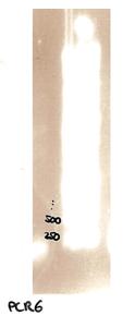 Agarose gel electrophoresis of PCR6 which shows that PCR6 is about 200bp Plate CMV and pDS7 colonies on Ampicillin-Agar
- colonies for plasmidextraction of CMV and pDS7 plated on Ampicillinplates
Plasmid Extraction of CMV and pDS7
- plasmidextraction of CMV (2,5ng/µl) and pDS7 (10ng/µl) the A260/A280 value was 1.333, which means that it was 90% Protein and only 10% DNA (should be 1,8); new plasmidextraction needed
new overnight cultures of CMV and pDS7 for a new plasmidextraction made
Agarose gel electrophoresis
-> Protocol (11 Agarose gel electrophoresis)
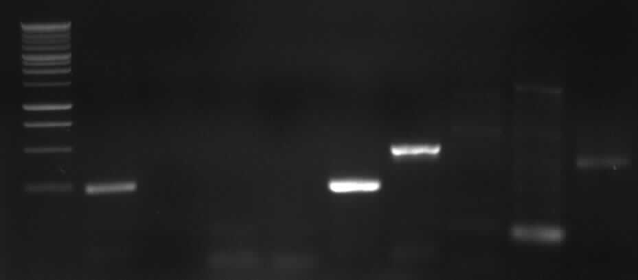
- Agarose gel electrophoresis of the restriction digest of PhiC31o and PCR 1 and 6
- the right bands found for PhiC31o (~2900,~2400,~250)
- the right band found for PCR1 (~450)
- no band found for PCR6; new electrophoresis needed with more DNA loaded

|

|
|
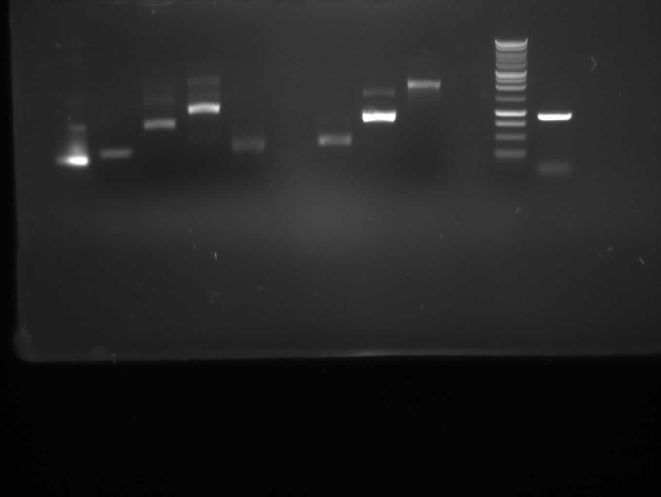
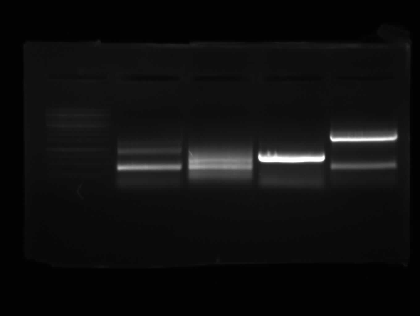
Agarose gel electrophoresis of (from left to right) PhiC31o, PCR1 and PCR6
|
Agarose gel electrophoresis of (from left to right) PhiC31o, PCR1 and PCR6 which shows that PCR1 is between 250 and 500 bp
|
- new agarose gel electrophoresis from PCR6 with 5µl DNA instead of 3µl (image not yet shown)
- the right band found for PCR6 (~200)
New overnight cultures of CMV and pDS7
- the overnight colonies didn't grow; new colonies (CMV and pDS7) picked from plate and inoculated in LB Ampicillin
PCR purification of PCR 1 and 6
-> Protocol (12 Gel extraction or PCR Clean up)
- DNA concentration of the PCR 1 and 6 products measured: PCR1: 410ng/µl (A260/A280=1.253) PCR6: 568ng/µl (A260/A280=1.275)
- PCR Purification with Promega Kit
-> PCR1: 230ng/µl (A260/A280=1.769)
-> PCR6: 37.5ng/µl (A260/A280=1.667)
8-18-2010
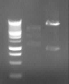 Agarose gel electrophoresis of (from left to right) CMV and pDS7 showing the right bands for pDS7 Plasmid Extraction of CMV and pDS7
-> Protocol (4 Plasmid extraction from cells)
- Plasmid extraction of CMV (97.5ng/µl; A260/A280=1.857) and pDS7 (212ng/µl; A260/A280=1.848)
Restriction digestion
-> Protocol (5 Restriction digestion)
- Restriction digestion of CMV (EcoR1 + Pst1; 10µl DNA, buffer H) and pDS7 (EcoR1 + Spe1; 5µl DNA, buffer B)
-> expected DNA bands: CMV: 2079bp (plasmid) + 654bp (Insert); pDS7: 7022bp + 1011bp
Agarose Gel electrophoresis of digested CMV and pDS7
-> Protocol (11 Agarorse gel electrophoresis)
-> right DNA bands for pDS7 (~7000bp, ~1000bp)
-> false DNA bands for CMV
- Starting PCR 2a and 2b (replication and mutagenesis of pDS7): 3 µl DNA and 50°C Annealing Temperatur (other same as 8-16-2010)
8-19-2010
Agarose gel electrophoresis of PCR 2a and 2b
-> Protocol (11 Agarose gel electrophoresis)
(150V, 30min)
|
| Agarose gel electrophoresis of (from left to right) PCR2a and PCR2b
|
-> the right bands for PCR2a (~300bp) and PCR2b (~700bp)
- New agarose gel electrophoresis with all of the PCR product for gel extraction (150V, 30min)
Gel extraction of the DNA from PCR2a and PCR2b
-> Protocol (12 Gel extraction or PCR Clean up)
- DNA concentration measured; problem with nanodrop as too low concentration; lyophille used to reduce volume
- DNA concentration measured again: PCR2a: 70ng/µl A260/A280=1.647; PCR2b: 45ng/µl A260/A280=1.5
PCR 3 (joining PCR of 2a and 2b)
- PCR3 (the joining PCR of PCR2a and 2b; Joining of the TEVrecogn-N-Degron-SF3 part) done: 1.3 µl of PCR2a and 4.7 µl of PCR2b makes 300ng of a 1:1 solution of both to be joined DNA parts. Annealing temperature: 50°C
-> Protocol (10 PCR with Pfu)
8-20-2010
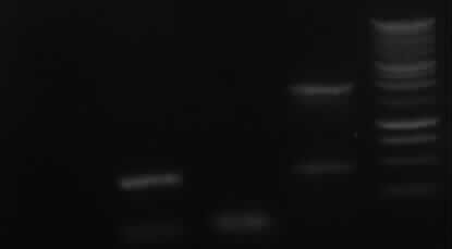
Agarose gel electrophoresis of PCR3
left column: marker; rightmost column: PCR3
-> Protocol: 11 Agarose gel electrophoresis (150V, 30min)
-> expected band: ~1000bp
-> false band: ~500bp
- probable reason: mini photometre was influenced by gel extraction chemicals, therefore it measured false DNA concentrations and false template masses were calculated
-> New 2a and 2b PCR
New PCR (2a and 2b)
-> Protocol: 10 PCR with Pfu
(see 8-18-2010, but 35,5µl water)
8-21-2010
weekend
8-22-2010
weekend
8-23-2010
Agarose gel electrophoresis of PCR 2b
-> Protocol: 11 Agarose gel electrophoresis
- expected band: 700bp
-> no band shown on gel -> new PCR 2b
PCR 2b
- start PCR 2b with PCR 2b from 8-13-10 as template ( 1:20 and 1:100 diluted; 1µl)
-> Protocol: 10 PCR with Pfu
- annealing temperature: 50°C; amount of water: 37,5µl
Agarose gel electrophoresis of PCR 2b 1:20 and 1:100
-> Protocol: 11 Agarose gel electrophoresis
- expected bands: each ~ 700bp
- false bands: ~ 200bp
-> new PCR with 2ng, 5ng, 10ng template pDS7
PCR 2b with 2ng, 5ng, 10ng template pDS7
- pDS7 1:100 diluted(-> 2,1 ng/µl)
Mixture:
|
| 2ng
| 5ng
| 10ng
|
| Primer
| 2*2,5µl (P5+P6)
| 2*2,5µl (P5+P6)
| 2*2,5µl (P5+P6)
|
| 10x Buffer Pfu
| 5µl
| 5µl
| 5µl
|
| dNTP Mix
| 1µl
| 1µl
| 1µl
|
| template
| pDS7 (dil.)
| 1µl
| 2,5µl
| 5µl
|
| Pfu Polymerase (3u/µl)
| 0,5µl
| 0,5µl
| 0,5µl
|
| DMSO
| 1,25µl
| 1,25µl
| 1,25µl
|
| H2O
| 33,25µl
| 30,25µl
| 25,25µl
|
| sum
|
|
|
|
-> Protocol: 10 PCR with Pfu
PCR 2a gel extraction
- Quiagen kit (QuiaexII)
-> Protocol: 14 QIAEX II gel extraction
Start 3 CMV overnight cultures
8-24-2010
agarose gel electrophoresis of PCR 2b
-> Protocol: 11 Agarose gel electrophoresis
 Agarose gel electrophoresis of (from left to right) PCR2b (2ng (cut out), 10ng, 5ng template) showing the right bands for 2ng, 5ng template - expected bands: right bands with 2ng and 5ng template (~700bp), no band with 10ng template
CMV plasmid extraction
-> Protocol: 4 Plasmid extraction from cells
Plasmid extractionof 3 different overnight cultures.
- results:
- 52,5 ng/µl A260/A280= 1.312
- 133 ng/µl A260/A280= 1.710
- 80 ng/µl A260/A280= 1.600
CMV restriction digestion
-> Protocol: 5 Restriction digestion
- CMV restriction digest: EcoRI, PstI, buffer H
- 19µl, H2O : 0µl
- 6µl, H2O : 9.5µl
- 12.5µl, H2O : 3µl
PCR 2b gel extraction
- PCR2b was gel extracted (with Qiagen gel extraction kit), 17.5 ng/µl a260/A280= 1.750
-> Protocol: 14 QIAEX II gel extraction
PCR 3 (fusion of 2a and 2b)
- PCR3: conducted again at 52°C annealing temperature
- 10.5 ng (from PCR2b) 0.6µl
- 4.5 ng (from PCR2a) 0.9µl (1:10 diluted)
| PCR2a
| 0.9 µl
|
| PCR2b
| 0.6 µl
|
| primer3
| 2.5 µl
|
| primer6
| 2.5 µl
|
| dNTPs
| 1 µl
|
| Pfu
| 0.5 µl
|
| 10xbuffer
| 5 µl
|
| H2O
| 37 µl
|
-> Protocol: 10 PCR with Pfu
agarose gel electrophoresis of CMV digestion
- agarose gel electrophoresis (150V, 25 min) of the CMV digestion
-> bands are wrong again ( ~ 1200bp, 2000bp)
8-25-2010
Agarose gel electrophorese of PCR 3
-> Protocol: 11 Agarose gel electrophoresis
- expected band: ~1000bp
- false band: ~400bp
Plasmid extraction of ccdB tet and ccdB strep
Plasmid extraction of pSB1C3 with BBa_P1010
-> Protocol: 4 Plasmid extraction from cells
- results:
| ccdB tet:
| 50ng/µl;
| A260/A280= 1,818
|
Plate ccdB amp, cam, tet
- plate ccdB with ampicilline, chloramphenicol, tetracycline resistance on LB agar with appropiate antibiotic.
Overnight culture of ccdB kan
- Overnight culture of ccdB with kanamycine resistance in LB medium with kanamycine
PCR 7a, 7b, 9, 10
->Protocol: 10 PCR with Pfu
| PCR nr.
| template
| concentration
| dilution
| primer
|
| 7a
| 190-6
| ~200ng/µl
| 1:100
| 13,14
|
| 7b
| 190-6
| ~200ng/µl
| 1:100
| 15,16
|
| 9
| eGFP
| 55ng/µl
| 1:25
| 20,21
|
| 10
| PhiC31o
| 20ng/µl
| 1:10
| 22,23
|
Mixture
| template (~2ng)
| 1µl
|
| Pfu
| 0,5µl
|
| Primer *2
| 2,5µl *2
|
| 10x buffer
| 5µl
|
| dNTP Mix
| 1µl
|
| H2O
| 37,5µl
|
| sum
| 50µl
|
Standard PCR; annealing temperature: 60°C
8-26-2010
Agarose gelelectrophoresis of PCR 7a, 7b, 9, 10
->Protocol: 11 Agarose gel electrophoresis
- 150V, 25min
| PCR nr.
| expected bands
| result
|
| 7a
| 850bp
| no band
|
| 7b
| 402bp
| false band (200bp)
|
| 9
| 808bp
| no band
|
| 10
| 1888bp
| no band
|
Plasmid extraction of ccdB kan
-> Protocol: 4 Plasmid extraction from cells
-result: concentration: 25ng/µl; A260/A280= 2,0
New PCR 7a, 7b, 9, 10
Mixture
| template (~2ng)
| 1µl
|
| Pfu
| 0,5µl
|
| Primer *2
| 2,5µl *2
|
| 10x buffer
| 5µl
|
| dNTP Mix
| 1µl
|
| DMSO
| 1,25µl
|
| H2O
| 36,25µl
|
| sum
| 50µl
|
Program:
gradient PCR, 42-69°C annealing temp.
->Protocol: 10 PCR with Pfu
Overnight culture of ccdB amp, tet, cam
Inoculate one colony each in 5ml medium with approptraite antibiotic.
8-27-2010
Agarose gel electrophoresis of PCR 7a, 7b, 9, 10
->Protocol: 11 Agarose gel electrophoresis
150V, 25min, 75mA
from left to right: 7a, 7b, 9, 10, Marker
| PCR nr.
| expected bands
| result
|
| 7a
| 850bp
| no band
|
| 7b
| 402bp
| right band (~400bp)+ false band (~150bp)
|
| 9
| 808bp
| false band (~200bp)
|
| 10
| 1888bp
| right band (~1900bp)+false band (~500bp)
|
Plasmid extraktion of ccdB amp, tet, cam
->Protocol: 4 Plasmid extraction from cells
results:
| Plasmid
| concentration
| A260/A280
|
| ccdB amp
| 57,5 ng/µl
| 1,917
|
| ccdB cam
| 70,0 ng/µl
| 1,867
|
| ccdB tet
| 50,0 ng/µl
| 1,818
|
New PCR 7a, 9
Mixture:
- 2ng template: see 26-8-10
- 4ng template: see 26-8-10, but 2µl template and 35,25µl water
-> Protocol: 10 PCR with Pfu
Restriction digestion of ccdB amp, kan, cam, tet
-> Protocol: 5 Restriction digestion
- only 90min 37°C incubation
- EcoRI, PstI, Buffer H
| template
| volume
| mass
|
| ccdB amp
| 16µl
| 930ng
|
| ccdB cam
| 14,3µl
| 1µg
|
| ccdB tet
| 16µl
| 800ng
|
| ccdB kan
| 16µl
| 400ng
|
Agarose gelelectrophoresis of PCR 7a, 9, ccdB restriction digestion
150v, 25min, 75mA
-> Protocol: 11 Agarose gel electrophoresis
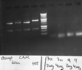
results:
- PCR7a, 9: false band at 200bp
- ccdB: each digestion leads to a right band with ~ 650bp
8-28-2010
weekend
8-29-2010
weekend
8-30-2010
New PCR 7a and 9
Mixture
| template (~4ng)
| 2µl
|
| Pfu
| 0,5µl
|
| Primer *2
| 2,5µl *2
|
| 10x buffer
| 5µl
|
| dNTP Mix
| 1µl
|
| DMSO
| 1,25µl
|
| H2O
| 35,25µl
|
| sum
| 50µl
|
-> Protocol: 10 PCR with Pfu
PCR program
PCR 7: Annealing Temperature 60°C - 25 x 1 min Annealing time and 5x 1,30 min Annealing time
PCR 9: Annealing Temperature 55°C - 25 x 1 min Annealing time and 5x 1,30 min Annealing time
Gel extraction of PCR 7b, 10
->Protocol: 14 QIAEX II gel extraction
results:
| PCR nr.
| concentration
| A260/A280
|
| 7b
| 10 ng/µl
| 2,0
|
| 10
| 17,5 ng/µl
| 1,4
|
Agarose gel electrophoresis of new PCR 7a, 9
-> Protocol: 11 Agarose gel electrophoresis
150V, 25min
- results:
- 7a: no band shown
- 9: false band (~200bp)
New PCR 3
-> Protocol: 10 PCR with Pfu
| PCR2a
| 0.9 µl
|
| PCR2b
| 0.6 µl
|
| dNTPs
| 1 µl
|
| Pfu
| 0.5 µl
|
| 10xbuffer
| 5 µl
|
| DMSO
| 1,25µl
|
| H2O
| 36,75 µl
|
| sum
| 45µl
|
- new method: standard PCR without primers (10 cycles, 56°C annealing temp.)
- then add 2,5µl of primer 3 and 6
- 30 cycles standard PCR (54°C annealing temp.)
8-31-2010
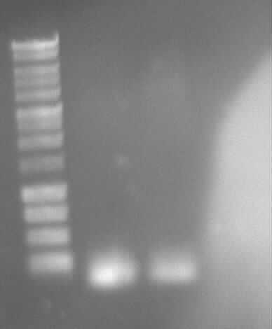
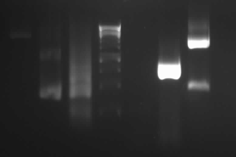
 Gel photo of (left to right) PCR4a(2.5ng template), PCR4a(5ng template),PCR4b(2.5ng template), PCR4b(5ng template), PCR3(Pfu), PCR3(Phusion) Agarose gel electrophoresis of PCR 4a, PCR4b, PCR3(Pfu), PCR3(Phusion)
-> Protocol: 11 Agarose gel electrophoresis
150V, 25min
- results:
- PCR4a(2.5ng template), PCR4a(5ng template),PCR4b(2.5ng template), PCR4b(5ng template), PCR3(Pfu): no band shown
- PCR3 (Phusion): right band (~1000bp)
New PCR PCR4a, PCR4b, PCR7a, PCR9
-> Protocol: 10 PCR with Pfu
PCR mixture for PCR4a, PCR4b
| template
| 37.25 µl (200ng)
|
| dNTPs
| 1 µl
|
| Pfu
| 0.5 µl
|
| 10xbuffer
| 5 µl
|
| DMSO
| 1,25µl
|
| sum
| 50µl
|
Standard PCR program with annealing temperature PCR4a: 51.1°C, PCR4b: 48.5°C.
-> Protocol: 15 PCR with Phusion
PCR mixture for PCR7a, PCR9
| template
| 4 µl (8ng)
|
| dNTPs
| 1 µl
|
| Phusion
| 0.5 µl
|
| 5xbuffer
| 10 µl
|
| DMSO
| 1,25µl
|
| H2O
| 28.25 µl
|
| sum
| 50µl
|
PCR program: Phu62
| 98°C
| 1 min
|
| 98°C
| 10 sec
|
| 62°C
| 20 sec
|
| 73°C
| 30 sec
|
| return to step 2 for 29 cycles
|
|
| 73°C
| 10 min
|
| 12°C
| forever
|
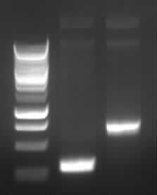
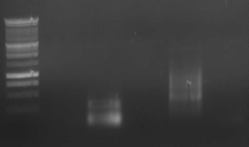
 Gel photo of (left to right) PCR3, ladder, PCR7a, empty, PCR9 Agarose gel electrophoresis of PCR 3, PCR7a, PCR9 for gel extraction
-> Protocol: 11 Agarose gel electrophoresis
120V, 30min
- results:
- PCR3; right band (~1000bp) and side-product
- PCR7a: no band
- PCR9: right band (~800bp) and side-product
Gel extraction of the DNA from PCR3 and PCR9
-> Protocol (12 Gel extraction or PCR Clean up)
results:
PCR 9: 22,5ng/µl; A260/A280=1,8
PCR 3: 22,5ng/µl; A260/A280=2,25
New PCR 4a, 4b, 7a with DreamTaq
-> Protocol: 16 PCR with DreamTaq
PCR mixture for PCR7a
| template
| 5 µl (10ng)
|
| dNTPs
| 5 µl
|
| DreamTaq
| 0.33 µl
|
| 10xbuffer
| 5 µl
|
| DMSO
| 1,25µl
|
| H2O
| 28.5 µl
|
| sum
| 50µl
|
Primers for PCR 7a: 13,14
Annealing temp: 60°C
PCR mixture for PCR4a,4b
| template
| 36,5 µl (180ng HeLa cDNA)
|
| dNTPs
| 5 µl
|
| DreamTaq
| 0.33 µl
|
| 10xbuffer
| 5 µl
|
| DMSO
| 1,25µl
|
| sum
| 50µl
|
PCR 4a
Primers for PCR 4a: 7,8
Primers for PCR 4b: 9,10
Annealing temp: 50°C
PCR program:
| 95°C
| 1 min
|
| 95°C
| 30 sec
|
| 50/60°C
| 30 sec
|
| 72°C
| 1 min (1kb/min)
|
| return to step 2 for 29 cycles
|
|
| 72°C
| 10 min
|
| 12°C
| forever
|
9-01-2010
Agarose gel electrophoresis of PCR4a, PCR4b, PCR7 (DreamTaq)
-> Protocol: 11 Agarose gel electrophoresis
150V, 25min
results: no product
New PCR 4a, 4b with DreamTaq, Pfu, with concentration gradient and touch-down PCR
-> Protocol: 16 PCR with DreamTaq; 10 PCR with Pfu
PCR mixture for DreamTaq
| Concentration
| Low
| Middle
| High
|
| template
| 5 µl (1:1000)
| 31.75µl (1:1000)
| 2µl (1:10)
|
| dNTPs
|
| 5 µl
|
|
| DreamTaq
|
| 0.33 µl
|
|
| 10xbuffer
|
| 5 µl
|
|
| DMSO
|
| 1,25µl
|
|
| H2O
| 26.75 µl
| 0
| 29.75
|
| sum
|
| 50µl
|
|
PCR mixture for Pfu
| Concentration
| Low
| Middle
| High
|
| template
| 5 µl (1:1000)
| 37.25µl (1:1000)
| 2µl (1:10)
|
| dNTPs
|
| 1 µl
|
|
| Pfu
|
| 0.5 µl
|
|
| 10xbuffer
|
| 5 µl
|
|
| DMSO
|
| 1,25µl
|
|
| H2O
| 32.25 µl
| 0
| 35.25 µl
|
| sum
|
| 50µl
|
|
Primers for PCR 4a: 7,8; PCR 4b: 9,10
-> Protocol: Thermal cycler program: Touch down
Agarose gel electrophoresis of PCR4a, PCR4b
-> Protocol: 11 Agarose gel electrophoresis
150V, 25min
from left to right: 4a: P1, P2, P3, D1, D2, D3; 4b: P1, P2, P3, D1, D2, D3
key:
"P"= PCR with Pfu
"D"= PCR with DreamTaq
"1"= low template concentration
"2"= middle template concentration
"3"= high template concentration
expected bands:
- 4a: 330bp -> P2 and P3 show right bands and "primer clouds"(?)
- 4b: 376bp -> P1, P2, P3 show right bands and "primer clouds" (?)
9-02-2010
Agarose gel electrophorese of PCR 4a P2, 4b P2
-> Protocol: 11 Agarose gel electrophoresis
- 120V, 45min, 1,5% Agarose gel
Agarose gel electrophoresis of (from left to right) PCR4aP2, Marker and PCR4bP2
- Cut out bands at ~~ 350bp and extract
PCR Agarose gel extraction
-> Protocol: 14 QIAEX II gel extraction
results:
| PCR nr.
| concentration
| A260/A280
|
| 4aP2
| 12,5 ng/µl
| 1,67
|
| 4bP2
| 72,5 ng/µl
| 1,53
|
New PCR 7a with Pfu and Phusion
template: 190-6, Primer 13,14
Mixture with Pfu
| template (~2ng)
| 1µl
|
| Pfu
| 0,5µl
|
| Primer *2
| 2,5µl *2
|
| 10x buffer
| 5µl
|
| dNTP Mix
| 1µl
|
| DMSO
| 1,25µl
|
| H2O
| 36,25µl
|
| sum
| 50µl
|
-> Protocol: 10 PCR with Pfu
PCR mixture with Phusion
| template (~2ng)
| 1,0 µl
|
| dNTPs
| 1 µl
|
| Phusion
| 0.5 µl
|
| 5xbuffer
| 10 µl
|
| DMSO
| 1,25µl
|
| H2O
| 31,25µl
|
| sum
| 50µl
|
-> Protocol: 15 PCR with Phusion
New PCR 4a, 4b with Pfu
-> Protocol: 10 PCR with Pfu
-> Protocol: Thermal cycler program: Touch down
Mixture see 9-1-10, twice 4a and 4b
Agarose gel electrophorese of PCR 4a, 4b, 7a, gel extracted 4a and 4b
-> Protocol: 11 Agarose gel electrophoresis
- 25min, 150V
from left to right: 4a*, 4a, 4b*, 4b, 7a Phusion, 7a Pfu, Ladder
-> result: 4b, 4b*: right bands (~330bp)
-remain: false bands/no band
from left to right: ladder, 4 columns pathway, 4a gelextr., 4b gelextr.
-> results: slight right bands for 4a and 4b, no "primer clouds" anymore.
9-03-2010
Overlapping PCR 5 with Pfu and Phusion
template: 4a, 4b Primer 7,10
Mixture with Pfu
| PCR4a (~15ng)
| 1.2µl
|
| PCR4b (~15ng)
| 2µl (1:10)
|
| Pfu
| 0.5µl
|
| Primer *2
| 2.5µl *2
|
| 10x buffer
| 5µl
|
| dNTP Mix
| 1µl
|
| DMSO
| 1,25µl
|
| H2O
| 34.05µl
|
| sum
| 50µl
|
-> Protocol: 10 PCR with Pfu
PCR program: standard PCR program for Pfu, Annealing temperature: 54°C
PCR mixture with Phusion
| PCR4a (~15ng)
| 1.2µl
|
| PCR4b (~15ng)
| 2µl (1:10)
|
| dNTPs
| 1 µl
|
| Phusion
| 0.5 µl
|
| 5xbuffer
| 10 µl
|
| DMSO
| 1,25µl
|
| H2O
| 29.05µl
|
| sum
| 50µl
|
-> Protocol: 15 PCR with Phusion
PCR program: standard PCR program for Phusion, Annealing temperature: 58°C
New PCR7a with Pfu
template: 190-6; Primer: 13,14
Mixture with Pfu
| template (~2ng)
| 1µl
|
| Pfu
| 0.5µl
|
| Primer *2
| 2.5µl *2
|
| 10x buffer
| 5µl
|
| dNTP Mix
| 1µl
|
| DMSO
| 1,25µl
|
| H2O
| 36.25µl
|
| sum
| 50µl
|
-> Protocol: 10 PCR with Pfu
PCR program: standard PCR program for Pfu, Gradient: 54.4°C, 57.8°C, 61.4°C, 65.0°C
-> results:
-7a: only "primer cluods"
-5 Pfu: no defined product (slurred?)
-5 Phusion: "primer clouds"
9-04-2010
weekend
9-05-2010
weekend
9-06-2010
charges for sequencing
| name
| 4a-7
| 4a-8
| 4b-9
| 4b-10
| 3-3
| 3-6
| 6-11
| 6-12
|
| DNA
| 4a; 2.4µl
| 4a; 2.4µl
| 4b; 0.5µl
| 4b; 0.5µl
| 3; 2µl
| 3; 2µl
| 6; 0.5µl
| 6; 0.5µl
|
| primer [1pmol/µl]
| 7; 3.2µl
| 8; 3.2µl
| 9; 3.2µl
| 10; 3.2µl
| 3; 3.2µl
| 6; 3.2µl
| 11; 3.2µl
| 12; 3.2µl
|
| Tris (10mM); pH 8.2
| 1.4µl
| 1.4µl
| 3.3µl
| 3.3µl
| 1.8µl
| 1.8µl
| 3.3µl
| 3.3µl
|
every charge 7µl
New overlapping PCR 5 with Pfu
template: 4a, 4b; 15ng
Primer 7,10
Mixture
| PCR4a (~7ng)
| 0.56µl
|
| PCR4b (~8ng)
| 1.1µl (1:10)
|
| Pfu
| 0.5µl
|
| Primer *2
| 2.5µl *2
|
| 10x buffer
| 5µl
|
| dNTP Mix
| 1µl
|
| DMSO
| 1,25µl
|
| H2O
| 36.6µl
|
| sum
| 50µl
|
-> Protocol: 10 PCR with Pfu
PCR program: standard PCR program for Pfu, Annealing temperature: 46.1°C,48.5°C, 51.1°C
-> results: "primer clouds"
New PCR 7a with Pfu
new dilution of 190-6; 1:100
| template[2ng/µl]
| 1µl
| 2µl
|
| pfu polymerase
| 0.5µl
| 0.5µl
|
| primer13
| 2.5µl
| 2.5µl
|
| primer14
| 2.5µl
| 2.5µl
|
| buffer
| 5µl
| 5µl
|
| dNTP Mix
| 1µl
| 1µl
|
| DMSO
| 1,25µl
| 1.25µl
|
| H2O
| 36.25µl
| 35.25
|
| sum
| 50µl
| 50µl
|
Agarose gel electrophoresis of 7a
-> results: "primer clouds"
charges for sequencing for PCR 1,7b and 9
| name
| 1-1
| 1-2
| 7b-15
| 7b-16
| 9-20
| 9-21
| 10-22
| 10-23
|
| DNA
| 1[1:10]; 1.74µl
| 1[1:10]; 1.74µl
| 7b; 3µl
| 7b; 3µl
| 9; 1.78µl
| 9; 1.78µl
| 10; 3.8µl
| 10; 3.8µl
|
| primer [1pmol/µl]
| 1; 3.2µl
| 2; 3.2µl
| 15; 3.2µl
| 16; 3.2µl
| 20; 3.2µl
| 21; 3.2µl
| 22; 3.2µl
| 23; 3.2µl
|
| Tris (10mM); pH 8.2
| 2.06µl
| 2.06µl
| 0.8µl
| 0.8µl
| 2.02µl
| 2.02µl
| 0µl
| 0µl
|
every charge 7µl
9-07-2010
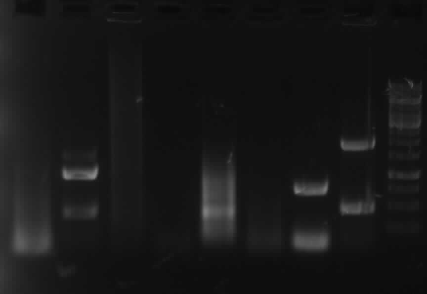
Agarose gel electrophorese of PCR gel extractions
150V, 25min
-> Protocol: 11 Agarose gel electrophoresis
-result:
from left to right: PCR 1,2a,2b,3,4a,/,7b,9,10,/,ladder,pathway
| PCR nr.
| 1
| 2a
| 2b
| 3
| 4a
| 7b
| 9
| 10
|
| expected band (bp)
| 492
| 332
| 772
| 1087
| 330
| 402
| 808
| 1888
|
| shown band(s)
| 550,200
| 300
| 750
| 1100
| 300
| 450
| 900,1500
| 1900
|
| clean charge
|
| x
| x
| ~x
| x
| x
|
| x
|
new PCR for PCR6
PCR mixture for PCR6
| Concentration
| Low
| High
|
| template
| 1 µl (1:100)
| 15µl (1:100)
|
| primer (11,12)
| 2.5 µl*2
|
|
| dNTPs
| 1 µl
|
|
| Pfu
| 0.5 µl
|
|
| 10xbuffer
| 5 µl
|
|
| DMSO
| 1,25µl
|
|
| H2O
| 36.25 µl
| 22.25 µl
|
| sum
| 50µl
|
|
-> Protocol: Thermal cycler program: Touch down, 62°C-52°C, 30 cycles by 55°C
9-08-2010
Restriction digestion of eGFP, PCR6, ccdBamp
 Gel photo of (left to right) PCR6(5ng/100ng) template as prepatation for ligation
-> Protocol 5 Restriction digestion
| eGFP
|
| SV40PA=PCR6
|
| ccdBamp
|
|
| DNA
| 5µl=300ng
| DNA
| 1µl=37,5ng
| DNA
| 2µl=115ng
|
| Buffer MC
| 2µl
| Buffer D
| 2µl
| Buffer H
| 2µl
|
| BSA 1:10
| 2µl
| BSA 1:10
| 2µl
| BSA 1:10
| 2µl
|
| EcoRI
| 0,5µl
| XbaI
| 0,5µl
| EcoRI
| 0,5µl
|
| SpeI
| 0,5µl
| PstI
| 0,5µl
| PstI
| 0,5µl
|
| H2O
| 13µl
| H2O
| 14µl
| H2O
| 13µl
|
| sum
| 23µl
| sum
| 20µl
| sum
| 20µl
|
1:30h 37°C, 20min 80°C
Agarose gel electrophoresis of PCR6
-> Protocol: 11 Agarose gel electrophoresis
25min, 150V
Ligation 1
| eGFP
| 14,4µL
| (144ng)
|
| SV40PA
| 4,8µL
| (48ng)
|
| ccdBamp
| 2,9µL
| (100ng)
|
| T4 Buffer 10x
| 3µL
|
|
| T4 Ligase
| 0,5µL
|
|
| H2O
| 4,4µL
|
|
| sum
| 30µL
|
|
Ligation at 22,5°C for 30 min, denaturation at 65°C for 10 min.
Concentration of DNA in Ligation 1:
Transformation
-> Protocol 18 competent cells2
The incubation time for the cells is here 1 hour.
9-09-2010
Agarose gel electrophoresis of PCR 1, 3, 4a, 5, 7a, 9
Agarose gel electrophoresis of PCR 1, 3, 4a, 5, 7a, 9
-> protocol 11 Agarose gel electrophoresis
results: "primer-clouds", PCR 9: no band
Plasmid extraction of ccdBcam, ccdBamp, Bak, CMV, PhiC31o
-> Protocol 4 Plasmid extraction from cells
| Plasmid
| concentration [ng/µL]
| A260/A280
|
| ccdBcam
| 22.5
| 1.0
|
| ccdBamp
| 42.5
| 1.2
|
| Bak
| 17.5
| 0.8
|
| CMV
| 10.0
| 0.7
|
| PhiC310
| 60
| 1.4
|
Eluated with H2O instead of the Eluation Buffer.
New PCR 1, 3, 4a, 5, 7a, 9, 10 with phusion
| PCRnr.
| 1
| 3
| 4a
| 4b
| 5
| 7a
| 9
| 10
|
| template
| pDS7 (1:100); 4µl
| 2a; 0.9µl+2b; 0.6µl
| Bak; 0.5µl
| Bak; 0.5µl
| 4a; 0.8µl+4b; 1µl
| 190-6 (1:100); 1µl
| eGFP (1:25); 2µl
| PhiC31o (1:10); 1µl
|
| primer [10pmol/µl]
| 1,2; 2.5µl
| 3,6; 2.5µl
| 7,8; 2.5µl
| 9,10; 2.5µl
| 7,10; 2.5µl
| 13,14; 2.5µl
| 20,21; 2.5µl
| 22,23; 2.5µl
|
| H2O
| 28µl
| 30.5µl
| 31.5µl
| 31.5µl
| 30.2µl
| 31µl
| 30µl
| 31µl
|
| Annealing temperature
| 51.5°C
| 53.1°C
| 53.1°C
| 51.5°C
| 53.1°C
| 57.5°C
| 61°C
| 57.5°C
|
+ in each assay:
dNTP mix: 1µl, 5x Phusion buffer: 10µl, DMSO:1.5µl, Phusion:0.5µl
sum: 50µl
Program: standard Phusion PCR, 29 cycles; annealing temperatures: see above
-> Protocol: 15 PCR with Phusion
[from left to right: PCR 1, 3, 4a, 5, 7a, 9, 10, Ladder]
PCR3, 9, 10 with right bands.
New PCR 1, 4a, 4b, 5, 7a with phusion
| PCRnr.
| 1
| 4a
| 4b
| 5
| 7a
|
| template
| pDS7 (1:10); 1µl
| Bak 1 ; 1µl
| Bak 1; 1µl
| 4a, 4b; 2 x 1.5µl
| pCT 190-6 (1:40); 1µl
|
| primer
| 1, 2; 2 x 2.5µl
| 7, 8; 2 x 2.5µl
| 9, 10; 2 x 2.5µl
| 7, 10; 2 x 2.5µl
| 13, 14; 2x 2.5µl
|
| H2O
| 31µl
| 31µl
| 31µl
| 30µl
| 30µl
|
| Annealing temperature
| 52°C
| 52°C
| 48°C
| 50°C
| 55°C
|
+ each assay with dNTP-mix (1µl), 5xBuffer (10µl, DMSO (1.5µl), Phusion (0.5µl)
-> sum: 50µl
-> Protocol Touch down 59 with phusion, 30 cycles with gradient appropriate for the annealing temperatures above.
9-10-2010
Agarose gel electrophoresis of PCR 1, 4a, 4b, 5, 7a
-> Protocol: 11 Agarose gel electrophoresis
[From left to right: Ladder, 1, 4a, 4b, 5, 7a]
Only 4a has been amplified successfully.
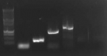
Agarose gel extraction of PCR 3, 4a, 5, 9, 10
from left to right: PCR 3, band ~700bp, PCR 4a, band ~300bp, PCR 5, bands ~550bp (5*), ~650bp (5), PCR 9, band ~800bp, PCR 10, band ~1900bp
results:
|
| PCR 3
| PCR 4a
| PCR 5
| PCR 5*
| PCR 9
| PCR 10
|
| concentration [ng/µl]
| 20
| 10
| 5
| 30
| 35
| 25
|
| A260/A280
| 1.6
| 1.333
| 2.0
| 2.0
| 1.750
| 2.0
|
-> Protocol 12 Gel extraction or PCR Clean up (Promega kit)
9-11-2010
weekend
9-12-2010
weekend
9-13-2010
charges for sequencing (retry of 6.9.)
| name
| 4a-7
| 4a-8
| 4b-9
| 4b-10
| 3-3
| 3-6
| 6-11
| 6-12
|
| DNA
| 4a; 2.4µl
| 4a; 2.4µl
| 4b; 0.5µl
| 4b; 0.5µl
| 3; 2µl
| 3; 2µl
| 6; 0.5µl
| 6; 0.5µl
|
| primer [1pmol/µl]
| 7; 3.2µl
| 8; 3.2µl
| 9; 3.2µl
| 10; 3.2µl
| 3; 3.2µl
| 6; 3.2µl
| 11; 3.2µl
| 12; 3.2µl
|
| Tris (10mM); pH 8.2
| 1.4µl
| 1.4µl
| 3.3µl
| 3.3µl
| 1.8µl
| 1.8µl
| 3.3µl
| 3.3µl
|
every charge 7µl
New PCR7a with Taq
template: p190-6
Primer 13,14
Mixture
| template (~4ng)
| 2µl (1:100)
|
| MasterMix for Taq
| 10µl
|
| Primer *2
| 1.5µl *2
|
| DMSO
| 0,5µl
|
| H2O
| 4.5µl
|
| sum
| 20µl
|
PCR program: touchdown PCR with Taq
1: 94°C 2'
|
| 2: 94°C 30"
|
| 3: 64°C/62°C/60°C/58°C/56°C/54° 30"
|
| 4: 72°C 2'
|
5: (for each temperature)repeat 2-4 2x
|
| 6: 94°C 30"
|
| 7: 58°C 30"
|
| 8: 72°C 2'
|
9: repeat 6-8 29x
|
| 10: 72°C 10'
|
| 11: 15°C break
|
-> Protocol: 21 PCR with Taq Mastermix
overnight culture inoculated of
- CMV (amp)
- ccdB (amp)
- ccdB (cam)
- pC31o (amp)
- Bak (amp)
New PCR7a with Phusion Hot Start
with 2ng and 4 ng template
| template
| 2ng
| 4ng
|
| H2O
| 54µl
| 53µl
|
| Buffer 5x
| 20µl
| 20µl
|
| Primer (13,14)
| 2*10µl
| 2*10µl
|
| DMSO
| 3µl
| 3µl
|
| Hot Start
| 1µl
| 1µl
|
total: 100µl each, divided into 5 charges
program:
- 98°C 30sec
- ----
- 98°C 10sec
- gradient: 50°C, 53°C, 56,6°C, 60,2°C, 64,5°C 30sec 30 cycles
- 72 15sec
- ----
- 72°C 5min
- 12°C forever
-> Protocol:20 PCR with Phusion Hot Start
9-14-2010
Agarose gelelectrophoresis of PCR 7a
-> Protocol: 11 Agarose gel electrophoresis
bands 1 to 5: 2ng of template DNA
bands 6 to 10: 4ng of template DNA
-> no bands
new PCR 7a and PCR 8 (without mutation)
with mastermix, without DMSO
| number:
| 7a diluted
| 7a undiluted
| 8
|
| mastermix
| 50µl
| 50µl
| 10µl
|
| primer
| 2*10µl
| 2*10µl
| 2*2µl
|
| H2O
| 29µl
| 29µl
| 5µl
|
| template
| 1µl 190-6 (1:100)
| 1µl 190-6
| 1µl 190-6 (1:100)
|
7a diluated and undiluated: divided into 3 charges
7a diluated: 1,2,3; 7a undiluated: 4,5,6
| 1
| 2
| 3
| 4
| 5
| 6
| 8
|
| 48°C
| 52°C
| 56,1°C
| 48°C
| 52°C
| 56,1°C
| 52°C
|
program:
94°C 2'
|
| 94°C 30"
|
| 48°C/52°C/56,1°C 30"
|
| 72°C 1,5'
|
(for each temperature)repeat 30x
|
| 72°C 5'
|
Plasmid extraction of ccdB (amp) and ccdB (cam)
-> Protocol: 4 Plasmid extraction from cells
results:
- ccdB(amp): 105ng/µl A260/280: 2,00
- cddB(cam): 102ng/µl A260/280: 1,952
9-15-2010
Agarose gelelectrophoresis of PCR 7a diluted &7a undiluted & "8"(without mutation)
-> Protocol: 11 Agarose gel electrophoresis
from left to right: 7a: 1:100 diluted: 48°C, 52°C, 56.1°C, undiluted: 48°C, 52°C, 56.1°C, 8 without Mutation
weak right band (with fuzz)for 7a undiluated with best result for 56°C
-> new PCR 7a and "8"
with mastermix Taq
-> Protocol: 21 PCR with Taq Mastermix
template: 190-6, 1:10 and 1:5
temp: 56°C and 58°C
| number
| 7a
| 7a
| "8"
| "8"
|
| Mastermix
| 20µl
| 20µl
| 20µl
| 20µl
|
| H2O
| 15µl
| 15µl
| 15µl
| 15µl
|
| Primer
| (13,14) 2*2µl
| (13,14) 2*2µl
| (13,16) 2*2µl
| (13,16) 2*2µl
|
| Template
| 190-6 (1:5) 1µl
| 190-6 (1:10) 1µl
| 190-6 (1:5) 1µl
| 190-6 (1:10) 1µl
|
2 charges each: one for 56°C and one for 58°C annealing temperature
program:
94°C 2'
|
| 94°C 30"
|
| 56°C/58°C 30"
|
| 72°C 1,5'
|
(for each temperature)repeat 30x
|
| 72°C 5'
|
Restriction Digestion of PCR 1 (17.8.), PCR 3 (10.9.), PCR 51 (10.9.), PCR 52 (10.9.), PCR 6 (17.8.), PCR 9 (10.9.), PCR 10 (30.8.) for ligation with vector
EcoR1 + Pst1 with Buffer H; 50ng DNA
|
| 1
| 3
| 51
| 52
| 6
| 9
| 10
|
| H2O
| 13µl
| 12,5µl
| 5µl
| 13µl
| 13,5µl
| 13,5µl
| 12,5µl
|
| Buffer H
| 2µl
| 2µl
| 2µl
| 2µl
| 2µl
| 2µl
| 2µl
|
| BSA (1:10)
| 2µl
| 2µl
| 2µl
| 2µl
| 2µl
| 2µl
| 2µl
|
| DNA
| 2µl
| 2,5µl
| 10µl
| 2µl
| 1,5µl
| 1,5µl
| 2,5µl
|
| EcoR1
| 0,5µl
| 0,5µl
| 0,5µl
| 0,5µl
| 0,5µl
| 0,5µl
| 0,5µl
|
| Pst1
| 0,5µl
| 0,5µl
| 0,5µl
| 0,5µl
| 0,5µl
| 0,5µl
| 0,5µl
|
-> Protocol: 5 Restriction digestion
Agarose gelelectrophoresis of PCR 7a 1-4 & "8" 1-4(without mutation)
-> Protocol: 11 Agarose gel electrophoresis
-> bad results
purification of restriction digestion
|
| concentration (ng/µl)
| A260/A280
| (supposed) lenght
|
| 1
| 20
| 1,000
| 492
|
| 3
| 5
| 1,000
| 1087
|
| 51
| 22,5
| 1,8
| 688
|
| 52
| 7,5
| 1,5
| 688
|
| 6
| 10
| 1,333
| 237
|
| 9
| 20
| 1,6
| 808
|
| 10
| 17,5
| 1,75
| 1888
|
-> Protocol: 12 Gel extraction or PCR Clean up (Promega kit)
Ligation with pSB1C3 (2072bp)
| for
| #
| backbone
| insert (µl)
| charge (µl)
| Buffer 10x (µl)
| H2O (µl)
|
| 100ng
| 1
| 4
| 7,12
| 20
| 2
| 6,38
|
| 50ng
| 3
| 2
| 31,48
| 40
| 4
| 2,02
|
| 100ng
| 51
| 4
| 8,85
| 20
| 2
| 4,65
|
| 100ng
| 52
| 4
| 26,56
| 40
| 4
| 4,94
|
| 100ng
| 6
| 4
| 6,86
| 20
| 2
| 13,36
|
| 100ng
| 9
| 4
| 11,7
| 20
| 2
| 1,8
|
| 100ng
| 10
| 4
| 31,24
| 40
| 4
| 4,26
|
plus 1 µl T4 ligase in each charge
-> Protocol: 22 Ligation
new PCR 7a & "8"
with mastermix Taq 56°C
|
| 7a-1
| 7a-2
| "8"-1
| "8"-2
|
| mastermix (µl)
| 10
| 10
| 10
| 10
|
| template
| 190-6 1µl
| 190-6 2µl
| 190-6(1:10) 1µl
| 190-6(1:10) 2µl
|
| primer
| 13&14 2*1µl
| 13&14 2*1µl
| 13&16 2*1µl
| 13&16 2*1µl
|
| H2O
| 7µl
| 6µl
| 7µl
| 6µl
|
sum: 20µl each
pcr-program:
94°C 2'
|
| 94°C 30"
|
| 56°C 30"
|
| 72°C 1,5'
|
(for each temperature)repeat 30x
|
| 72°C 5'
|
| 12°C forever
|
-> Protocol: 21 PCR with Taq Mastermix
9-16-2010
Agarose gelelectrophoresis of PCR 7a-1, PCR 7a-2, PCR 8-1, 8-2 and of all three PCR 6 we ever made (in order to control whether we have extracted the right fragment (237) or just the primerdimers)
-> Protocol: 11 Agarose gel electrophoresis
From left to right: 62, 61, 6, Ladder, 7a-1, 7a-2, 8-2, 8-1
- results:
- 6-1 and 6-2: ?
- 6: ok
- 7a-1,7a-2,"8"-1,"8"-2: primerdimer-problem -> we ordered new primers!
transformation of the ligations 1,3,51, 52, 6, 9, 10
-> Protocol:3 Transformation
->plated on plates with chloramphenicol
again PCR 1,3,5,6,9,10 in order to increase the profit (Ausbeute) for ligation
|
| 1
| 3
| 5
| 6
| 9
| 10
|
| template
| prev TRE(1:30) 1µl
| 2a(1:5) 1µl + 2b 0,5µl
| 4a 0,5µl + 4b 0,5µl
| SV40PA (pcDNA3)(1:50) 1µl
| eGFP(1:5) 1µl
| PhiC310(1:6) 1µl
|
| primer
| 1&2 2*2,5µl
| 3&6 2*2,5µl
| 7&10 2*2,5µl
| 11&12 2*2,5µl
| 20&21 2*2,5µl
| 22&23 2*2,5µl
|
| Tm
| 54°C
| 50°C
| 49°C
| 56°C
| 58°C
| 58°C
|
| mastermix (µl)
| 25
| 25
| 25
| 25
| 25
| 25
|
| DMSO
| 1,25µl
| 1,25µl
| 1,25µl
| 1,25µl
| 1,25µl
| 1,25µl
|
| H2O (µl)
| 17,75
| 17,25
| 17,75
| 17,75
| 17,75
| 17,75
|
pcr-program:
94°C 2'
|
| 94°C 30"
|
| Tm (see above) 30"
|
| 72°C 1,5'
|
(for each temperature)repeat 30x
|
| 72°C 5'
|
-> Protocol: 21 PCR with Taq Mastermix
9-17-2010
Analysis of the transformation from yesterday
Colonies on plates when 100µl or pellet plated:
|
| 1
| 3
| 5
| 6
| 9
| 10
|
| pellet
| no
| yes
| yes
| yes
| yes
| yes
| yes
|
| 100µl
| no
| yes
| yes
| yes
| yes
| yes
| yes
|
again: transformation of ligation 1
->Protocol: 3 Transformation
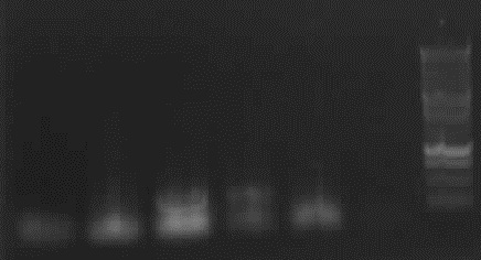
Agarose gelelectrophoresis of yesterday's PCR 1,3,5,6,9,10(without mutation)
-> Protocol: 11 Agarose gel electrophoresis
From left to right: PCR1, PCR3, PCR5, Ladder, PCR6, PCR9, PCR10
-> right band for PCR 3,5,6,9,10; no band for PCR 1
From left to right: Ladder, PCR3, PCR5, PCR9, PCR10
Bands took: 32: Band short over 1000bp, 31: Band short under 1000bp (probably 32 right), 5 the highest band, 9 upper band, 10 upper band
new PCR 1,5,6
|
| 1
| 5
| 6
|
| template
| pTRE Rev 0,5µl
| 4a 1,5µl + 4b 1,5µl
| pcDNA3 0,5µl
|
| primer
| 1&2 2*2,5µl
| 7&10 2*2,5µl
| 11&12 2*2,5µl
|
| buffer 10x
| 5µl
| 5µl
| 5µl
|
| dNTPs
| 1µl
| 1µl
| 1µl
|
| Pfu
| 0,5µl
| 0,5µl
| 0,5µl
|
| H2O
| 26,75 µl
| 24,25 µl
| 26,75 µl
|
| T<sub<m</sub>
| 51°C
| 50°C
| 55°C
|
-> Protocol:
pcr-program: standard Pfu 10 PCR with Pfu
Restriction digestion of PCR products, ccdB Plasmids and Biobricks for the 3A Method
| Name
| H2O
| Buffer (each 2µl)
| BSA (1:10)
| DNA Volume
| DNA Mass
| Enzyms (2*0.5µl)
|
| ccdBa
| 25 µl
| H
| 2µl
| 20µl
| ~600ng
| E+P
|
| ccdBa
| 25µl
| H
| 2µl
| 20µl
| ~700ng
| E+P
|
| 1
| 43µl
| MC
| 2µl
| 2µl
| ~500ng
| E+S
|
| 3
| 25µl
| B
| 2µl
| 20µl
| ~200ng
| X+P
|
| Primer 18+19
| 25µl
| B
| 2µl
| 10µl+10µl
| ?
| X+P
|
| 51
| 25µl
| MC
| 2µl
| 20µl
| ~100ng
| E+S
|
| 52
| 25µl
| MC
| 2µl
| 20µl
| ~600ng
| E+S
|
| 6
| 35µl
| B
| 2µl
| 10µl
| ~400ng
| X+P
|
| 9
| 25µl
| MC
| 2µl
| 20µl
| ~700ng
| E+S
|
| 10
| 25µl
| MC
| 2µl
| 20µl
| ~400ng
| E+S
|
| eGFP
| 35µl
| MC
| 2µl
| 10µl
| ~550ng
| E+S
|
| CMV1
| 35µl
| H
| 2µl
| 10µl
| ~530ng
| E+P
|
-> Protocol:5 Restriction digestion
Colony PCR of Ligations 3, 51, 52, 6, 9, 10 and PCR of CMV1 (to test if right)
10 1-4, 9 1-4, 6 1-4, 51 1-4, 52 1-4, 3 1-4 Colonies picked and put in following Mix:
| PCR Mastermix
| 10µl
|
| Biobrick Primer F
| 1.5µl
|
| Biobrick Primer R
| 1.5µl
|
| H2O
| 7µl
|
CMV1:
| PCR Mastermix
| 10µl
|
| Biobrick Primer F
| 1.5µl
|
| Biobrick Primer R
| 1.5µl
|
| Template
| 2µl
|
| H2O
| 5µl
|
-> Protocol: 21 PCR with Taq Mastermix
Agarose Gel electrophoresis of 3, 51, 52, 6, 9, 10 and CMV1 (to test if right)
-> Protocol: 11 Agarose gel electrophoresis
Above: From left to right: 3.1, 3.2, 3.3, 3.4, 51.1, 51.2, 51.3, 51.4, Ladder, 52.1, 52.2, 52.3, 52.4
Below: From left to right: 6.1, 6.2, 6.3, 6.4, 9.1, 9.2, 9.3, 9.4, Ladder, 10.1, 10.2, 10.3, 10.4, CMV1
-> right bands for 6.3 and 6.4, CMV1 ~1200bp (we think that this is right, as Biobrick sequenzing information indicates that it isn't 654bp but about 1200bp)
PCR clean up of PCR Gel extraction 31, 32, 5, 9, 10, PCR Product 6 and digestion PCR1, Primer 18+19, R51, R52, PCR6, PCR9, PCR10, PCR3
-> Protocol: 12 Gel extraction or PCR Clean up (Promega kit)
9-18-2010
weekend
9-19-2010
weekend
9-20-2010
Dephosphorylation of linearized ccdB amp and ccdB cam
Add 1µl TSAP to digested vectors.
Incutation: 37°C 15min; Inhibition: 74°C 15min
Ligation and 3A-Assemblies
Jump-or-Die Ligations: 1a, 1b (51),1b (52), 2, 3
Cut'N'Survive Ligations: 1a, 2b,
Biobricks for both Systems: CMV, PCR1 (tet-on-promoter)
Mixtures:
- 3A Assemblies (everything except CMV, PCR1):
Inserts: each 8µl; ccdB vector: 1µl; T4 Ligase: 0.5µl; T4 Ligase Buffer: 2µl; H2O: 1µl
- Biobricks:
Insert: 10µl; ccdB vector: 1µl; T4 Ligase: 0.5µl; T4 Ligase Buffer: 2µl; H2O: 7µl
Incubation: 2:30h 16°C; Inhibition: 10 min 65°C
->Protocols: 22 Ligation, 13 3A Method for Biobrick assembly
Colony PCR of PCR product ligations with pSB1C3
Mixture:
PCR Mastermix:130µl ; PrimerF:19,5µl ; PrimerR:19,5µl ; H2O:91µl
-> Protocol: 21 PCR with Taq Mastermix
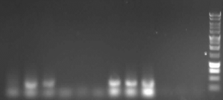
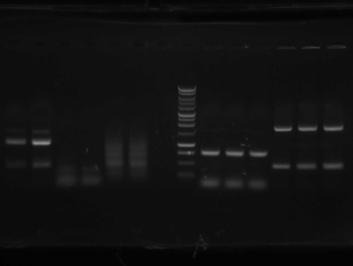
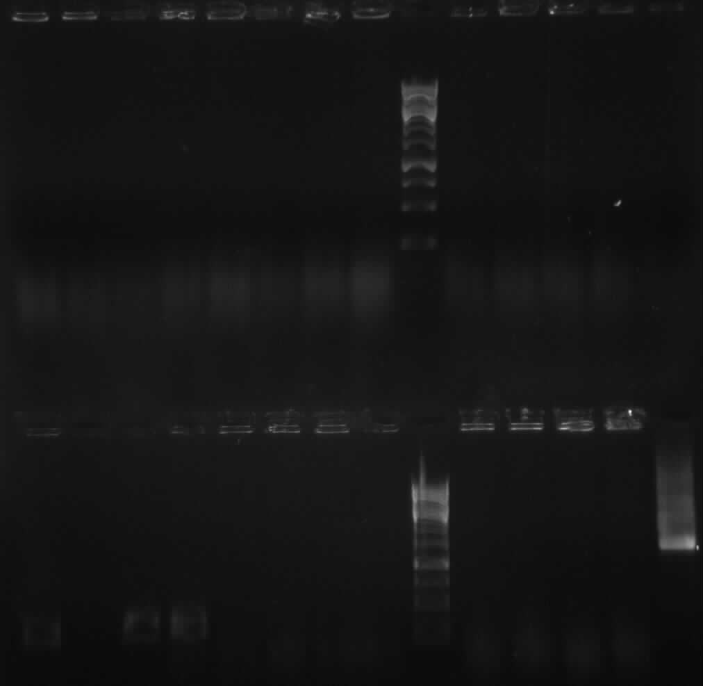
Agarose Gel Electrophoresis of Colony PCRs
-> Protocol: 11 Agarose gel electrophoresis
above from left to right: pathway/ladder/3.5/3.6/3.7/3.8/51.5/51.6/51.7/51.8/52.5/52.6/52.7/52.8
below from left to right: ladder/6.5/6.6/6.7/6.8/9.5/9.6/9.7/9.8/10.5/10.6/10.7/10.8
9-21-2010
Agarose Gel Electrophoresis of PCRs 1, 5, 6, and Colony PCR 3.8, 51.7, 52.6, 9.7, 10.5, 10.6
-> Bad results, probably too much cells in PCR mix
-> Protocol: 11 Agarose gel electrophoresis
Colony PCR of 3.8, 51.7, 52.6, 9.7, 10.5, 10.6 again and of the 3A Ligations of yesterday as well as the Ligation of PCR1 and CMV with pSB1C3
PCR Master Mix: 60µl
Primer F: 9µl
Primer R: 9µl
H2O: 42µl
->22charges à 5µl
-> Protocol:21 PCR with Taq Mastermix
PCR 7a with new Primers
| template 190-6 (1:10)
| 1µl
|
| Master Mix
| 25µl
|
| Primer 13k (short)
| 3,75µl
|
| Primer 14
| 3,75µl
|
| H2O
| 16,5µl
|
sum: 50µl
same with Primer 13l (long)
->Protocol: 21 PCR with Taq Mastermix
Agarose Gel Electrophoresis of Colony PCRs and PCR 7a
different ladder:
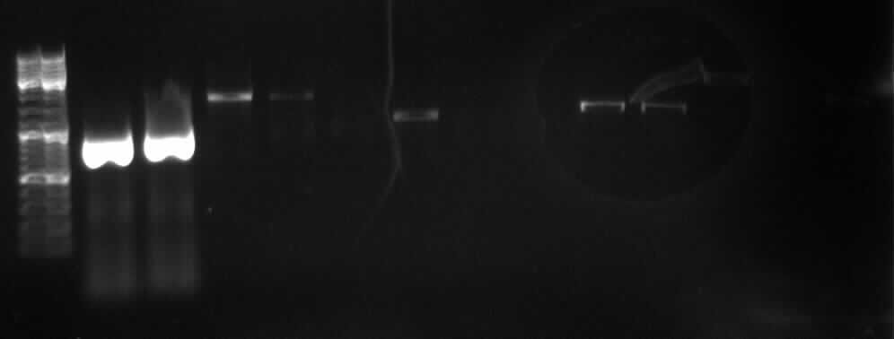
key: JD=Jump-or-Die; CS=Cut'N'Survive; first number=Ligation number (see schedule); second number=colonie number (marked on the plate)
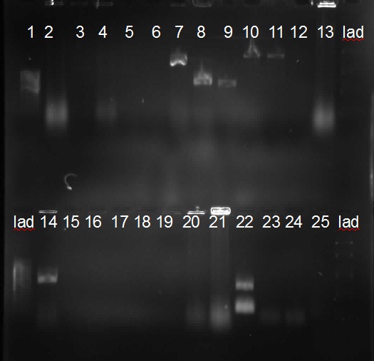
from left to right:7ak(short primer), 7al(long primer), JD.1a.1, JD.1a.2,JD.1b51.1, JD.1b52.2, JD.1b52.1, JD.1b52.2, JD.2.1, JD.2.2, JD.31.1, JD.31.2, CS.1a.1, CS.1a.2
verified products:7a (~800bp); JD.2.1; JD.2.2(each ~1000bp);JD.31.1(~2000bp); CS.1a.2(~1500bp)
no bands shown: CS.2b.1, CS.2b.2, PCR1.1, PCR1.CMV,3.8, 51.7, 52.6, 9.7, 10.5, 10.6
-> Protocol: 11 Agarose gel electrophoresis
9-22-2010
Inoculate Colonies
inoculated in 4ml LB-medium with appropriate antibiotic
Plated residual transformated E. colis (9-20-10) (where we had few colonies on plates)
Colony PCRs
for 22*5µl charges:
| PCR Mastermix
| 55µl
|
| Primer F
| 8,25µl
|
| Primer R
| 8,25µl
|
| H2O
| 38,5µl
|
| sum
| 110µl
|
-> Protocol: 21 PCR with Taq Mastermix
Annealing temperature: 53°C
PCR Purification of 7a
charges 7ak and 7al had not been labeled, so they were renamed.
results:
7a1: 135ng/µl; A260/A280=1.7
7a2: 133ng/µl; A260/A280=1.7
PCR8 with Phusion Hot Start II
-> Protocol: 20 PCR with Phusion Hot Start
| template 7a 1:100
| 3µl ~4ng (850bp)
|
| template 7b 1:10
| 2µl ~2ng (402bp)
|
| Buffer 5X
| 10µl
|
| Phusion
| 0.5µl
|
| H2O
| 27µl
|
| Primer 13
| 2.5µl
|
| Primer 16
| 2.55µl
|
| DMSO)
| 1.5µl
|
| dNTP mix
| 1µl
|
Program: annealing temperature: 63°C, elongation time: 40sec
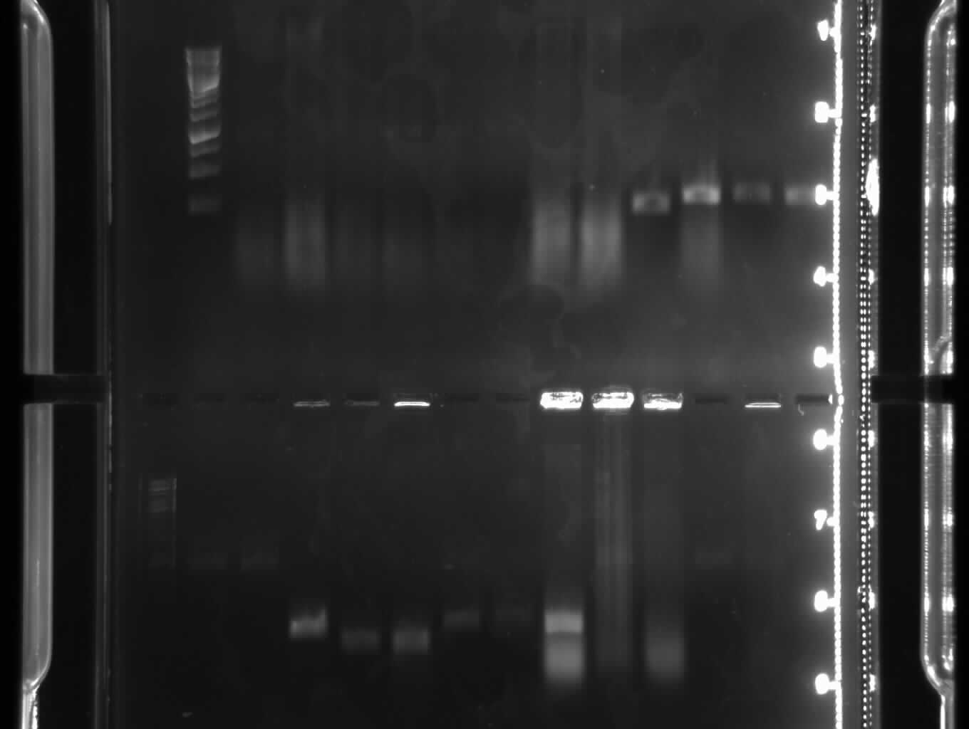
Agarose gel electrophoresis of Colony PCRs and PCR8
new ladder: fermentas FastRuler (TM) Middle Range DNA Ladder http://www.fermentas.com/en/products/all/dna-electrophoresis/fastruler-dna-ladders/sm111-fastruler-middle
some results:
PCR 8 (lanes 23, 24): no band
Colony PCRs CS.2b.10, PCR1cam.3, PCR1cam.4, CMVcam.4, CMVcam.4, JD.1b51.10 (lanes 7,8,9,10,11,14): show right bands
Plasmid extraction of "over-day" cultures ligation-7(=BB4),-8(=BB4),-9(=BB5),-12(=BB9) and pSB1C3
->Protocol: 4 Plasmid extraction from cells
concentrations (ng/µl - A260/280):
- BB4(7):57,5 - 1,643
- BB4(8):15 - 1,5
- BB5(9):173 - 1,816
- BB7(12):55,0 - 1,571
- pSB1C3: 12,5 - 1,250
new PCR 7b in order to get better results
| Master Mix
| 25µl
|
| H2O
| 16,5µl
|
| Primer 15
| 3,75µl
|
| Primer 16
| 3,75µl
|
| template 190-6 (1:10)
| 1µl
|
overnight-cultures
inoculated of: CS2bK10=BB11; PCP1K3+4=BB1; CMV-C-K3+4; JD1b51K10=BB2; pSB1C3
9-23-2010
Restriction digestion of BB5 for the 3A Method
| Name
| H2O
| Buffer D
| BSA (1:10)
| DNA Volume
| DNA Mass
| Enzyms (2*0.5µl)
|
| BB5
| 11 µl
| 2µl
| 2µl
| 4µl
| ~500ng
| X+P
|
sum: 20µl
-> Protocol:5 Restriction digestion
Plasmid extraction of overnight cultures BB2, BB11, CMV in cam and pSB1C3
->Protocol: 4 Plasmid extraction from cells
concentrations (ng/µl - A260/280):
- BB2:232 - 1,603
- BB11:170 - 1,889
- CMV in cam:272 - 1,703
- pSB1C3: 290 - 1,758
The pSB1C3 colony was pink (produced a red pigment or something like that, so we make a new transformation in order to extract only pSB1C3 and not some other vectors.
Agarose gelelectrophoresis of PCR 7b and pathway restriction digestions
-> Protocol: 11 Agarose gel electrophoresis
From left to right: PCR7b, pathway (3x),ladder (5000,2000,850,400,100bp)
result: PCR 7b shows right band with ~400bp
PCR clean up of PCR 7b
-> Protocol: 12 Gel extraction or PCR Clean up (Promega kit)
result: 90ng/µl; A260/A280= 1,895
Restriction digestion of BB2 and BB11
| Name
| H2O
| Buffer D
| BSA (1:10)
| DNA Volume
| DNA Mass
| Enzyms (2*0.5µl)
|
| BB2
| 10 µl
| 2µl
| 2µl
| 5µl
| ~1.2mg
| X+P
|
sum: 20µl
| Name
| H2O
| Buffer D
| BSA (1:10)
| DNA Volume
| DNA Mass
| Enzyms (2*0.5µl)
|
| BB11
| 7 µl
| 2µl
| 2µl
| 8µl
| ~1.3mg
| X+P
|
sum: 20µl
-> Protocol 5 Restriction digestion
Ligation of BB1 and BB6
-> Protocol: 22 Ligation
| Name
| H2O
| T4 Ligase-Buffer
| PCR1
| Primer 18+19
| ccdBAmp
| T4 Ligase
|
| BB1
| 9.5 µl
| 2µl
| 5µl
| 2µl
| 1µl
| 0.5µl
|
| Name
| H2O
| T4 Ligase-Buffer
| CMV E+S
| BB5
| ccdBCam
| T4 Ligase
|
| BB6
| 6.5 µl
| 2µl
| 8µl
| 2µl
| 1µl
| 0.5µl
|
PCR 8 with 7b(new) plus 7a-2 with Phusion Hot Start II; Primer 16 plus 13k and 13l
| Buffer 5x
| 10µl
|
| H2O
| 27µl
|
| dNTPs
| 1µl
|
| Primer 16
| 2,5µl
|
| Primer 13k short
| 2,5µl
|
| DMSO
| 1,5µl
|
| template 7a(1:100)(850bp)
| 3µl -> 4ng
|
| template 7b(new)(1:100)(402bp)
| 2µl -> 2ng
|
| Phusion Hot Start II
| 0,5µl
|
- ->same with Primer 13l long<-
Overnight culture of PCR1 in cam- Vector
new inoculation of PCR1 in cam, Colony 3 in 5ml LB+cam
 From left to right: PCR8k,PCR8l,ladder (5000,2000,850,400,100bp) Transformation of pSB1C3, BB1, BB6
New transformation of pSB1C3 from Spring distribution because the old colony was pink.
Transformation of BB1 and BB6.
BB1 plated on amp-Agar, BB6 and pSB1C3 plated on cam-Agar
-> Protocol: 19 Transformation (Sina Science Services)
Agarose gelelectrophoresis of PCR8 (long and short primer 13)
PCR8k=short primer 13, PCR8l=long primer 13
-> Protocol: 11 Agarose gel electrophoresis
result: PCR 8k and 8l shows right band with ~1200bp
PCR purification of PCR 8
-> Protocol: 12 Gel extraction or PCR Clean up (Promega kit)
result: ~70ng/µl; A260/A280=1.75
9-24-2010
New "over-day" culture of pSB1C3
Inoculate one culture from the ligation yesterday in 4ml LB+cam
Ligation of BB72
Mixture:
Inserts: each 5µl; ccdB vector: 1µl; T4 Ligase: 0.5µl; T4 Ligase Buffer: 2µl; H2O: 7µl
Incubation: 5h 18°C; Inhibition: 20 min 65°C
->Protocols: 22 Ligation, 13 3A Method for Biobrick assembly
Colony PCRs of BB1 and BB6
2 Colonys were picked from each plate -> 4 charges
Mixture for 4 charges with 5µl:
| PCR Mastermix 2x
| 10µl
|
| Primer F
| 1.5µl
|
| Primer R
| 1.5µl
|
| H2O
| 7µl
|
| sum
| 20µl
|
+ Colony
 from left to right: 1 1, 1 2, 6 1 and 6 2-> Protocol: 21 PCR with Taq Mastermix
Annealing temperature: 53°C, standard protocol
Plasmid extraction of new PCR1 in cam vector
-> Protocol: 4 Plasmid extraction from cells
results:
| concentration
| 82.5ng/µl
|
| A260/A280
| 1.737
|
Agarose Gelelektrophoresis of 11, 12, 61 and 62
Plasmid extraction of pSB1C3 in cam vector
-> Protocol: 4 Plasmid extraction from cells
results:
| concentration
| 22.5ng/µl
|
| A260/A280
| 1.5
|
9-25-2010
weekend
9-26-2010
weekend
9-27-2010
Colony PCR of BB72, BB6 and BB1 plus a negativ charge as reference
| PCR Mastermix
| 17.5µl
|
| Primer forward
| 2.625µl
|
| Primer revers
| 2.7µl
|
| H2O
| 12.1µl
|
| sum
| 35µl = 5µl per charge
|
-> Protocol 24 Colony PCR
Ligation of BB10
| CMV© E+S
| 5µl
|
| PCR 8l X+P
| 5µl
|
| ccdBamp
| 1µl
|
| T4 Ligase-Buffer
| 2µl
|
| T4 Ligase
| 0.5µl
|
| H2O
| 6.5µl
|
| sum
| 20µl
|
-> Protocol 22 Ligation
Ligation of BB001(PCR1 in ©), BB017(PCR5 in ©), BB018(PCR6 in ©) and BB021(PCR9 in ©)
| ccdB
| 1µl
|
| BB001/017/018/021
| 5µl
|
| T4 Ligase-Buffer
| 2µl
|
| T4 Ligase
| 0.5µl
|
| H2O
| 11.5µl
|
| sum
| 20µl
|
-> Protocol 22 Ligation
Restriction digestion of PCR32, 8l, 10 and Primer 18+19 with E+P
|
| template
| H2O
| BSA [1:10]
| Buffer H
| EcoRI
| PstI
|
| PCR32
| 10µl
| 5µl
| 2µl
| 2µl
| 0.5µl
| 0.5µl
|
| PCR8l
| 10µl
| 5µl
| 2µl
| 2µl
| 0.5µl
| 0.5µl
|
| Primer 18+19
| 2x7.5µl
| --
| 2µl
| 2µl
| 0.5µl
| 0.5µl
|
| PCR10
| 10µl
| 5µl
| 2µl
| 2µl
| 0.5µl
| 0.5µl
|
-> Protocol 5 Restriction digestion
9-28-2010
Plating BB017, BB021, BB018, BB001, BB72, BB10
| Amp
| BB72
| BB10
|
|
|
| Cam
| BB001
| BB017
| BB018
| BB021
|
Colony PCR BB1, BB6, BB72 with DreamTaq (each 2 colonies)
-> Protocol: 16 PCR with DreamTaq
PCR mixture for PCR7a
| template
| pick up colony from plate
|
| primers*2
| 0.25µl*2
|
| dNTPs
| 0.5µl
|
| DreamTaq
| 0.05µl
|
| 10xbuffer
| 0.5µl
|
| H2O
| 3.45µl
|
| sum
| 5µl
|
Primers for colony PCR: BBseqF, BBseqR
Annealing temperature: 53°C
Ligation of BB2, BB019, BB020, BB016, BB022
|
| BB2
| BB019
| BB020
| BB016
| BB022
| Volume
|
| PCR
| 52
| 8
|
| 32
| 10
| each 5µl
|
| PCR
| 6
|
|
|
|
| each 5µl
|
| Primer
|
|
| 18+19
|
|
| each 5µl
|
| ccdBamp
| 1µl
|
|
|
|
|
|
| ccdBcam
|
| 1µl
| 1µl
| 1µl
| 1µl
|
|
| T4 Ligase-Buffer
| 2µl
| 2µl
| 2µl
| 2µl
| 2µl
|
|
| T4 Ligase
| 0.5µl
| 0.5µl
| 0.5µl
| 0.5µl
| 0.5µl
|
|
| H2O
| 6.5µl
| 11.4µl
| 11.4µl
| 11.4µl
| 11.4µl
|
|
| sum
| 20µl
| 20µl
| 20µl
| 20µl
| 20µl
|
|
9-29-2010
plates BB72, BB017, BB10, BB021, BB001, BB018: nothing grown -> thrown into trash
colony-PCR of BB2/8, BB019, BB016, BB020, BB022 with Taq
->protocol: PCR with Taq
| H2O
| 6,7µl
|
| Buffer 10x
| 1µl
|
| MgCl2
| 1µl
|
| dNTPs
| 0,2µl
|
| Primer forw. seq f
| 0,5µl
|
| Primer rev. seq r
| 0,5µl
|
| Taq
| 0,1µl
|
template: pick colony
sum: 10µl; we made 100µl ->10 charges (2 colonies of each plate)
tube numbers:
| 1
| 2
| 3
| 4
| 5
| 6
| 7
| 8
| 9
| 10
|
| BB2/BB8 - 1
| BB2/BB8 - 2
| BB016 - 1
| BB016 - 2
| BB019 - 1
| BB019 - 2
| BB020 - 1
| BB020 - 2
| BB022 - 1
| BB022 - 2
|
PCR-program: taq53
agarose-gelelektrophoresis -> no bands visible
new plates made of BB1, BB4, BB6, BB7
Ligations BB72, BB017 (mit PCR51), BB10, BB021, BB001, BB018
|
| PCR (5 µl each)
| PCR (5µl each)
| ccdB(amp)
| ccdB©
| Buffer
| T4 Ligase
| H2O
|
| BB72O
| 1
| 32
| 1µl
|
| 2µl
| 0,5µl
| 6,5µl
|
| BB017
| 51
|
|
| 1µl
| 2µl
| 0,5µl
| 11,5µl
|
| BB10
| CMV©
| PCR8
| 1µl
|
| 2µl
| 0,5µl
| 6,5µl
|
| BB021
| 9
|
|
| 1µl
| 2µl
| 0,5µl
| 11,5µl
|
| BB001
| 1
|
|
| 1µl
| 2µl
| 0,5µl
| 11,5µl
|
| BB018
| 6
|
|
| 1µl
| 2µl
| 0,5µl
| 11,5µl
|
restriction degestions
of PCR6, PCR52, PCR32, PCR1, ccdB(amp), ccdB(cam)
| template
| digestion with
| volume
|
| PCR 6
| Xba1 & Pst1
| I. 10µl II. 3µl
|
| PCR52
| EcoR1 & Pst1
| 20µl
|
| PCR32
| Xba1 & Pst1
| 10µl
|
| PCR1
| EcoR1 & Spe1
| 2µl
|
| ccdB(amp)
| EcoR1 & Pst1
| 20µl
|
| cddB(cam)
| EcoR1 & Pst1
| 20µl
|
->protocol: 5 Restriction digestion
colony-PCR of BB4/c3, BB6/c7, BB72/c5, BB1/c5, (BB11 as control) with Taq
->protocol: PCR with Taq
Mastermix:
| H2O
| 32µl
|
| Buffer 10x
| 5µl
|
| MgCl2
| 5µl
|
| dNTPs
| 1µl
|
| Primer forw. seq f
| 2,5µl
|
| Primer rev. seq r
| 2,5µl
|
| Taq
| 0,5µl
|
| DMSO
| 1,5µl
|
| sum
| 50µl
|
->10µl for each charge
template: pick colony; BB11: 2ng
tube numbers:
| 1
| 2
| 3
| 4
| 5
|
| BB4-c3
| BB6-c7
| BB72-c5
| BB1-c5
| BB11
|
PCR program:TA = 53°C; elongation time=1min
9-30-2010
Colony PCR of BB2/BB8, BB6 K7&K8 and test PCR of BB11 with new dilutions and primers
| Colony/BB11
| 2.5µl
|
| H2O
| 32µl
|
| dNTP
| 1µl
|
| Primer F
| 2.5µl
|
| Primer R
| 2.5µl
|
| MgCl2
| 5µl
|
| DMSO
| 1.5µl
|
| Buffer 10x (with KCl)
| 5µl
|
| Taq Polymerase
| 0.5µl
|
| sum
| 56µl
|
-> Protocol 24 Colony PCR
PCR purification of the digestions of 6I, 6II, 52, 32, 1
|
| concentration [ng/µl]
| A260/A280
|
| 6II
| 32.5
| 1.444
|
| ccdBamp
| 0.0
| ---
|
| ccdBcam
| 5.0
| 1.000
|
| 6I
| 0.0
| ---
|
| 52
| 20.0
| 1.143
|
| 32
| 27.5
| 1.57
|
| 1
| 25
| 1.667
|
-> Protocol 12 Gel extraction or PCR Clean up (Promega kit)
Transformation of BB018, BB72, BB001, BB017, BB10 and BB021
-> Protocol 19 Transformation SSS
Plasmidextraction of the following overnight cultures: BB020(K1), BB018(K5), BB6(K5), BB022(K1), BB016(K1), BB019(K1), BB2(K1) and BB018(K3)
|
| concentration [ng/µl]
| A260/A280
|
| BB020(K1)
| 163
| 1.711
|
| BB018(K5)
| 92.5
| 1.85
|
| BB6(K5)
| 120
| 1.778
|
| BB022(K1)
| 105
| 1.75
|
| BB016(K1)
| 85
| 1.889
|
| BB019(K1)
| 105
| 1.909
|
| BB2(K1)
| 190
| 1.81
|
| BB018
| 110
| 1.833
|
-> Protocol 4 Plasmid extraction from cells
10-01-2010
new test PCR of BB5 and BB11 with 20µl and 50µl volume
Master Mix
| H2O
| 67µl
|
| dNTP
| 2µl
|
| Primer F
| 5µl
|
| Primer R
| 5µl
|
| MgCl2
| 10µl
|
| Buffer 10x (with KCl)
| 10µl
|
| Taq Polymerase
| 1µl
|
| sum
| 100µl
|
template: BB5/50µl: 5ng, 20µl: 1.5ng; BB11/20µl: 3ng
->protocol: PCR with Taq
from left to right: ladder, BB5/50µl, BB5/20µl, BB11/20µl. BB5/50µl and BB11/20µl show the right band.
10-02-2010
weekend
10-03-2010
weekend
10-04-2010
sequencing
cycle, clean and run
|
| CMV (c)
| PCR 1 (c)
| BB4 7
| BB4 8
| BB018K5
| BB018K3
| BB6K5
| BB2K1
|
| template
| 1µl
| 2µl
| 3µl
| 3,8µl
| 2µl
| 2µl
| 2µl
| 1µl
|
| primer (1:100)
| 3,2 µl
| 3,2 µl
| 3,2 µl
| 3,2 µl
| 3,2 µl
| 3,2 µl
| 3,2 µl
| 3,2 µl
|
| 10mM TrispH 8,5
| 2,8 µl
| 1,8 µl
| 0,8 µl
| 0 µl
| 1,8 µl
| 1,8 µl
| 1,8 µl
| 1,8 µl
|
| name of tube
| 028 FIR
| 001 F/R
| 4-7 F/R
| 4-8 F/R
| 018-5 F/R
| 018-3 F/R
| 6-5 F/R
| 2-1 F/R
|
PCR
|
| 019
| 022
| 016
| 020
|
| template (1:100 = 2ng)
| 2µl
| 2µl
| 2,5µl
| 1,5µl
|
| Primer F
| 1µl
| 1µl
| 1µl
| 1µl
|
| Primer R
| 1µl
| 1µl
| 1µl
| 1µl
|
| dNTPs
| 0,4µl
| 0,4µl
| 0,4µl
| 0,4µl
|
| Taq
| 0,2µl
| 0,2µl
| 0,2µl
| 0,2µl
|
| Buffer
| 2µl
| 2µl
| 2µl
| 2µl
|
| MgCl2
| 2µl
| 2µl
| 2µl
| 2µl
|
| H2O
| 11,4µl
| 11,4µl
| 10,9µl
| 11,
|
| colour of tube
| orange
| rose
| green
| blue
|
10-05-2010
Colony PCRs of BB6, BB4, BB021, BB022, CMV©, BB001, BB016, BB1, BB252, BB72, BB017, BB018, BB019, BB020
agarose gel electrophoresis
schedule of the agarose gel electrophoresis:
| gel 1
| BB6K9
| BB6K10
| BB6K11
| BBK12
| BB6K13
| BB4K5
| ladder
| BB4K6
| BB4K7
|
|
| BB021K14
| BB021K13
| BB021K12
| BB021K11
| BB022K18
| BB022K19
| ladder
| BB022K17
| BB022K16
|
| gel 2
| BB022K12
| BB022K11
| CMV©K4
| CMV©K5
| BB001K5
| BB001K6
| BB001K7
| BB001K8
| ladder
|
|
| BB016K17
| BB1K16
| BB1K15
| BB1K8
| BB1K7
| BB252K1
| BB252K2
| BB252K3
| ladder
|
| gel 3
| BB017K14
| BB017K13
| BB017K12
| BB017K11
| BB018K17
| BB018K16
| BB018K15
| BB018K14
| ladder
|
|
| BB019K6
| BB019K5
| BB019K4
| ladder
| BB019K3
| BB020K7
| BB020K8
| BB020K6
| BB020K5
|
| gel 1
| BB4K8
| BB4K9
| BB4K10
| BB021K10
| BB021K15
|
|
|
|
|
|
| BB022K15
| BB022K14
| BB022K13
| ---
| ---
|
|
|
|
|
| gel 2
| BB001K9
| BB001K10
| BB016K11
| BB016K12
| BB016K13
| BB016K14
| BB016K15
| BB016K16
| ---
|
|
| ---
| BB252K4
| BB252K5
| BB72K12
| BB72K11
| BB72K10
| BB72K9
| BB017K16
| BB017K15
|
| gel 3
| BB018K13
| BB018K12
| BB018K11
| BB018K10
| BB018K9
|
|
|
|
|
|
| BB020K4
| BB020K3
| ---
| ---
|
|
|
|
|
|
Gelbilder und Auswertung einfügen
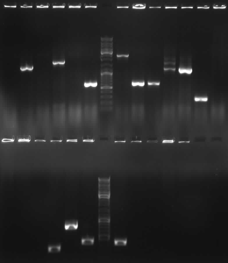
overnight cultures of BB6K3, BB4K10, CMV©K4, BB001K5, BB001K9, BB016K11, BB1K8, BB252K3, BB72, BB017K14, BB018K10, BB019K6
10-06-2010
Plasmidextraction of the following overnight cultures: BB6(K13), BB4(K13), CMVc (K4), BB001(K5), BB001(K9), BB016(K11), BB1(K8), BB2(K3), BB7(K10), BB017(K14), BB018(K10) and BB019(K6)
|
| concentration [ng/µl]
| A260/A280
|
| BB6(K13)
| 265
| 1.8
|
| BB4(K10)
| 40
| 2.0
|
| CMVc(K4)
| 97.5
| 1.8
|
| BB001(K5)
| 30
| 1.7
|
| BB001(K9)
| 17.5
| 1.7
|
| BB016(K11)
| 45
| 1.7
|
| BB1(K8)
| 57.5
| 1.9
|
| BB2(K3)
| 60
| 1.8
|
| BB7(K10)
| 30
| 1.7
|
| BB017(K14)
| 12.5
| 1.5
|
| BB018(K10)
| 35
| 1.8
|
| BB019(K6)
| 37.5
| 1.5
|
-> Protocol 4 Plasmid extraction from cells
10-07-2010
colony PCR of BB021, BB022 with Taq (53°C)
| Primer fw
| 0.5 µl
|
| Primer rev
| 0.5 µl
|
| dNTPs
| 0.2 µl
|
| Taq
| 0.1 µl
|
| 10xbuffer
| 1 µl
|
| MgCl2
| 1 µl
|
| H2O
| 6.7 µl
|
10-08-2010
plasmide extractions from 10-06-2010 were put in a lyophilizer and redissolved in less water (10-20 µl)
|
| concentration [ng/µl]
| A260/A280
|
| BB4(K10)
| 258
| 1,981
|
| BB001(K5)
| 325
| 1.94
|
| BB001(K9)
| 205
| 1.907
|
| BB7(K10)
| 170
| 1.744
|
| BB017(K14)
| 215
| 1.955
|
| BB018(K10)
| 500
| 1.88
|
| BB019(K6)
| 293
| 1.95
|
10-09-2010
Plasmid extraction BB018K5, BB018K6, BB021K17 and BB021K24
concentration of these samples to be measured on Monday
-> Protocol 4 Plasmid extraction from cells
10-10-2010
weekend
10-11-2010
Plasmid extraction BB018K5, BB018K6, BB021K24 and BB021K17 from 10-09-2010
|
| concentration [ng/µl]
| A260/A280
|
| BB018K5
| 55
| 1.571
|
| BB018K6
| 92,5
| 1.762
|
| BB021K24
| 27.5
| 1.833
|
| BB021K17
| 37.5
| 1.508
|
Colony PCR BB01, BB018, BB6, BB252
| stripe 1
| BB001K5
| K6
| K7
| K8
| K9
| K9
| K10
| BB252 K2
|
| stripe 2
| BB252 K3
| K4
| K5
| BB6 K9
| K10
| K11
| K12
| K13
|
| stripe 3
| BB018 K9
| K10
| K11
| K12
| K13
| K14
| K15
| K16
|
| single sample
| BB018 K17
|
gel picture colony PCR
foto einfügen!
Sequencing 8,9 and 10 (cycle, clean and run)
|
| BB001K9 for
| BB001K9 rev
| BB4K16 for
| BB4K16 rev
| BB001K5 for
| BB001K5 rev
| BB252K3
| BB252K3
|
| template
| 1µl
| 1µl
| 1µl
| 1µl
| 0,5µl
| 0,5µl
| 3,8µl
| 3,8µl
|
| primer (1:100)
| 3,2 µl
| 3,2 µl
| 3,2 µl
| 3,2 µl
| 3,2 µl
| 3,2 µl
| 3,2 µl
| 3,2 µl
|
| 10mM TrispH 8,5
| 2,8 µl
| 2,8 µl
| 2,8 µl
| 2,8 µl
| 3,3 µl
| 3,3 µl
| 0 µl
| 0 µl
|
| name of tube
| 31-F
| 31-R
| 44-F
| 44-R
| 24-F
| 24-R
| 32-F
| 32-R
|
|
| BB72K10 for
| BB72K10 rev
| BB017K14 for
| BB017K14 rev
| BB018K10 for
| BB018K10 rev
| BB019K6 for
| BB019K6 rev
|
| template
| 1,5µl
| 1,5µl
| 1µl
| 1µl
| 0,5µl
| 0,5µl
| 0,8µl
| 0,8µl
|
| primer (1:100)
| 3,2 µl
| 3,2 µl
| 3,2 µl
| 3,2 µl
| 3,2 µl
| 3,2 µl
| 3,2 µl
| 3,2 µl
|
| 10mM TrispH 8,5
| 2,3 µl
| 2,3 µl
| 2,8 µl
| 2,8 µl
| 3,3 µl
| 3,3 µl
| 3 µl
| 3 µl
|
| name of tube
| 14-F
| 14-R
| 21-F
| 21-R
| 13-F
| 13-R
| 34-F
| 34-R
|
|
| CMV for
| CMV rev
| BB018K5 for
| BB018K5 rev
| BB018K6 for
| BB018K6 rev
| BB021K17
| BB021K17
|
| template
| 2µl
| 2µl
| 3,8µl
| 3,8µl
| 2µl
| 2µl
| 3,8µl
| 3,8µl
|
| primer (1:100)
| 3,2 µl
| 3,2 µl
| 3,2 µl
| 3,2 µl
| 3,2 µl
| 3,2 µl
| 3,2 µl
| 3,2 µl
|
| 10mM TrispH 8,5
| 1,8 µl
| 1,8 µl
| 0 µl
| 0 µl
| 1,8 µl
| 1,8 µl
| 0 µl
| 0 µl
|
| name of tube
| CMV-F
| CMV-R
| 185-F
| 185-R
| 186-F
| 186-R
| 2117-F
| 2117-R
|
ligation BB020 BB022, Bb6 and BB52
-> Protocol 19 Transformation SSS
|
| BB020
| BB022
| BB6
| BB52
|
| insert (5µl)
| 18+19
| PCR 10
| CMV
| PCR5<sub<2</sub>
|
| insert (5µl)
| -
| -
| BB5
| PCR6
|
| ccdB (1µl)
| c
| c
| c
| a
|
| buffer
| 2µl
| 2µl
| 2µl
| 2µl
|
| T4-ligase
| 0,5µl
| 0,5µl
| 0,5µl
| 0,5µl
|
| H2O
| 11,5µl
| 11,5µl
| 6,5µl
| 6,5µl
|
PCR to test self-made taq
| template eGFP
| 1µl
|
| Primer 20,21
| each 1µl
|
| buffer (10x)
| 2µl
|
| MgCl
| 2µl
|
| dNTPs
| 0,2µl
|
| taq
| 0,3µl to 0,7µl (0,1µl steps)
|
| H2O
| fill to 20 µl
|
anealing temperature 58°C, elongation time 30
|
 |
 "
"


