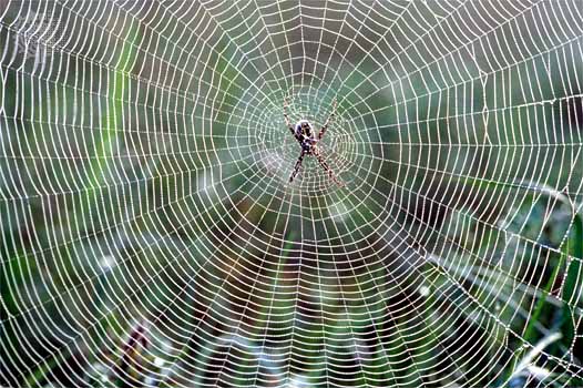
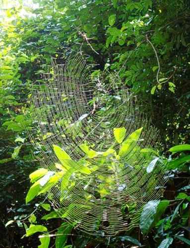
| Spider silk is comparable in strength to carbon fibres
|
| Highly structured at the nanometre scale – not good for synthetic materials
|
| Repetitive structures- GXG motif
|
| Glycine rich segments – hard and soft segments alternating
|
| Hard= hydrogen bonding cross-linked crystallites (polyalanine) forming an amorphic beta sheet structure,
|
| Soft= flexibility (Glycine rich)
|
| Major protein from Nephila clavipes – MaSP1 tandem variants of
|
| A GQG GYG GLG SQG A GRG GLG GQG A GA6GGx
|
| MaSP2 also has a repetitive structure – difference soft segment contains proline containing pentamers: The consensus repeat is _GPGGY GPGQQ.3GPSGPGS A8. Similar structure to Elastin – elastic properties of drag-line by the folding of pentamer structure.
|
| In the spider – silk in 3 phases
|
| 1) Extremely viscous (withstand shear forces inside spider),
|
| 2) Liquid crystallite lower viscosity (near exit duct/glycine rich may be involved),
|
| 3) Insoluble fibre (result of dehydration and drawing).
|
| MaSP1 and MaSP2 – Drag line
|
| MaSP1-Auxilary
|
| MaSP2- Glue silk only
|
| Neither- Cocoon silk
|
| Super contraction associated with pentamer motif when wet: low visco-elasticity
|
| Mimic natural proteins or simplify – Mimic structural significance still uncertain for some sequences
|
| DPB1- Optimised for B.subtilis
|
| B.subtilis potential host as simple secretion system compared to yeast. Secretion has advantages over expression in E.coli however; insufficient proportion of protein was secreted by yeast.
|
| Fahnestock, S. R., Yao, Z., & Bedzyk, L. a. (2000). Microbial production of spider silk pr}oteins. Journal of biotechnology, 74(2), 105-19. Retrieved from http://www.ncbi.nlm.nih.gov/pubmed/11763501.
|
| Self assembly of Spider silk proteins is controlled by a pH sensitive relay.
|
| A drop to pH 6.3 and shearing forces are believes to result in the hard form of spider silk.
|
| Miniature spidroins (a few repeats 4repCT) form metre long fibres irrespective of pH.
|
| Introduction of N-terminal domain from MASP1 (Euprosthenops australis)
|
| to mini spidroins allows self assembly at pH 6.3 and delays aggregation above pH7.
|
| Relay- like mechanism N terminal regulates assembly. pH 8 NT-mini spidroins for long firbes over days at pH 6 they form rapidly.
|
| C-terminal domain promotes Silk formation by ordering the repetitive segments.
|
| Askarieh, G. et al., 2010. Self-assembly of spider silk proteins is controlled by a pH-sensitive relay. Nature, 465(7295), 236-238. Available at: http://www.nature.com/doifinder/10.1038/nature08962.
|


 "
"