Team:Washington/Gram Positive/Design
From 2010.igem.org
Contents |
Making CapD a Better Anthrax Treatment
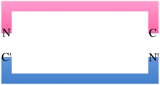
There are two main obstacles limiting natural CapD as an Anthrax therapeutic. First, natural CapD is a difficult to express dimer, requiring an auto-cleavage to activate. Second, CapD is a better transpeptidase than poly-γ-D-glutamate hydrolase, limiting its Anthrax decapsulating potential. To solve the first problem, we created a circular permuted, monomeric version of CapD that is easy to express and quantify. To improve hydrolysis, we used FoldIt, a computational toolbox, to design active site mutations aimed to increase hydrolysis over transpeptidation.
Circular Permutation of CapD
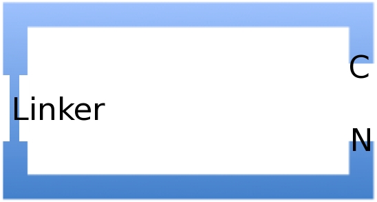
When natural CapD is first translated, the key catalytic threonine residue is buried in the active site, inaccessible to poly-γ-D-glutamate. After auto-cleavage, this critical threonine becomes the new N terminus, which can take its place in the active site. By reordering the protein so the threonine is the first residue, and putting a FoldIt designed linker between the natural N and C terminus, we make a circular permutation of CapD that we named CapD_CP. CapD_CP is a monomer, historically easier to purify and more stable than dimers.
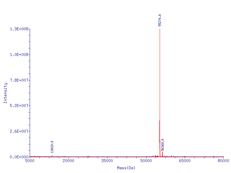
Since the first residue of any nascent protein must be methionine, we rely on E. Coli’s naturally occurring methionine aminopeptidase to remove the first methionine, making CapD_CP catalytically active. The removal of the first methionine has been verified via mass spectrometry.
The advantage of CapD_CP
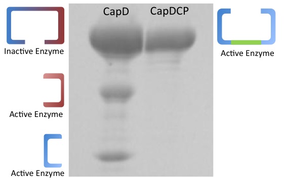
In addition to the fact that CapD_CP is easy to express, it has one more crucial advantage over CapD. When purifying CapD_CP, given our mass spec data, we can assume a massive majority of the purified CapD_CP is functional. CapD, however, is a much more ambiguous case, as illustrated by our protein gel results. The lower two bands of CapD correspond to the two subunits of cleaved CapD, which are active. The upper band may be uncleaved, therefore unfunctional, CapD, or dimeric, active CapD that simply did not denature, or a combination of both. Because of this, its difficult to quantify how much active protein is in a solution of purified CapD, making assaying activity a nightmare. For this reason, we made our active site mutations to CapD_CP.
Using FoldIt to Make CapD_CP a Better Hydrolase
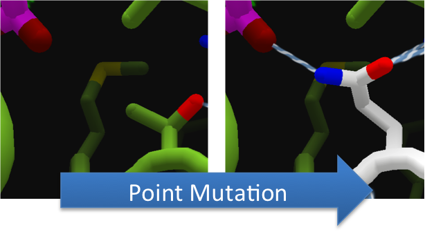
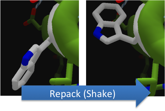
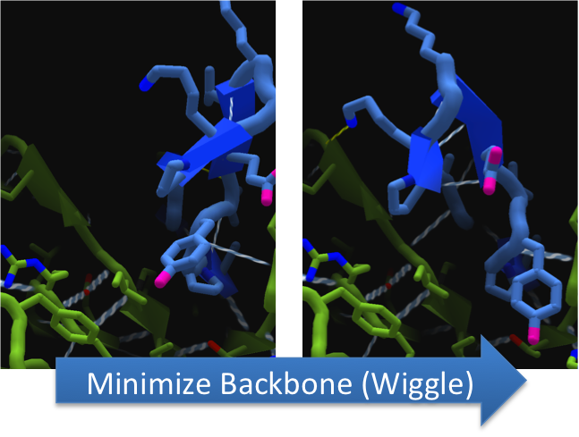
To increase the hydrolytic ability of CapD_CP, we made point mutations to the active site. We focused our attention on two types of mutations. First, we created point mutations that hydrogen bond to a modeled transition state of our substrate in an attempt to lower the activation energy, making hydrolysis more favorable. Second, we mutated the active site into a more open and polar area in an attempt to increase the ease with which water can enter and participate in a hydrolysis reaction.To make these point mutations, we used a computer program named FoldIt to predict how changes in protein structure and composition will affect protein stability. FoldIt provides a 3D representation of a protein's crystal structure that can be manipulated. Manipulation functions include point mutations, insertions, deletions, repacking of side chains (rotamer optimization), and backbone movement, which FoldIt then assesses for stability. This allows the user to quickly interact with a protein and easily predict how mutations will affect a protein.
A more in depth explanation of FoldIt here.
 "
"

