Team:Newcastle/solution

From 2010.igem.org

Revision as of 15:49, 27 October 2010 by Shethharsh08 (Talk | contribs)

iGEM Homepage Newcastle University BacillaFilla Homepage Image Map

Contents

Overview

BacillaFilla repairs concrete by producing a mixture of calcium carbonate, levan glue and filamentous cells in the cracks. Once we have applied BacillaFilla spores onto the concrete surface, they will start germinating in the presence of media. Once the cells have germinated, they will start to swarm down the crack. At the bottom of the crack when they reach a high density, they will use subtilin quorum sensing to activate concrete repair. BacillaFilla repairs concrete by 3 different processes:

  1. Some of the cells with produce calcium carbonate crystals,
  2. Some of the cells will become filamentous thereby acting as reinforcing fibres in the crack and
  3. All the cells will produce Levans glue which acts as a binding agent and at the same time it fills up the whole crack.


Therefore the mixture of all the three elements together will make a strong repair.

The industrial process of BacillaFilla starts from the batch bioreactor where the cells are grown at optimal conditions. The cells are then made to undergo sporulation and the spores are then transferred into storage containers. Spores don't require any nutrition or constant care and therefore they are ideal for long term storage and transportation. The storage containers will then be transported to the concrete repair site where they can be attached to a hand operated sprayer which will enable the personnel to spray spores along with the media onto the concrete surface. Once landed onto the concrete surface in the presence of media, the spores will germinate and will initiate concrete repair.

Our Initial Research

Parts Submitted to the Registry

  1. CaCO3/Urease
  2. Filamentous cells
  3. End of crack & signalling system
  4. Swarming
  5. Non-target-environment kill switch
  6. Glue


Testing and Characterisation

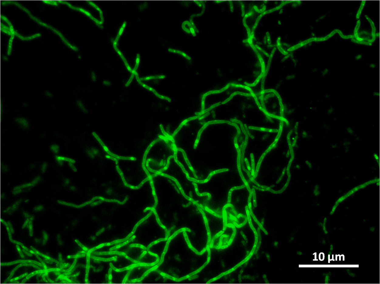
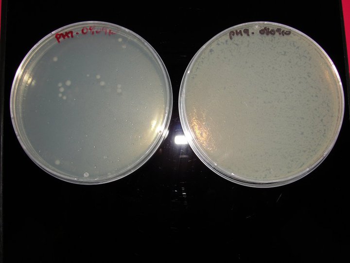
Newcastle filamentous gfp expt1.jpg Newcastle philrach levan.jpg Newcastle pH test.jpg

(Images above 1-3 left to right)

  1. yneA Characterisation
  2. Levansucrose glue test
  3. Alkalinity resistance

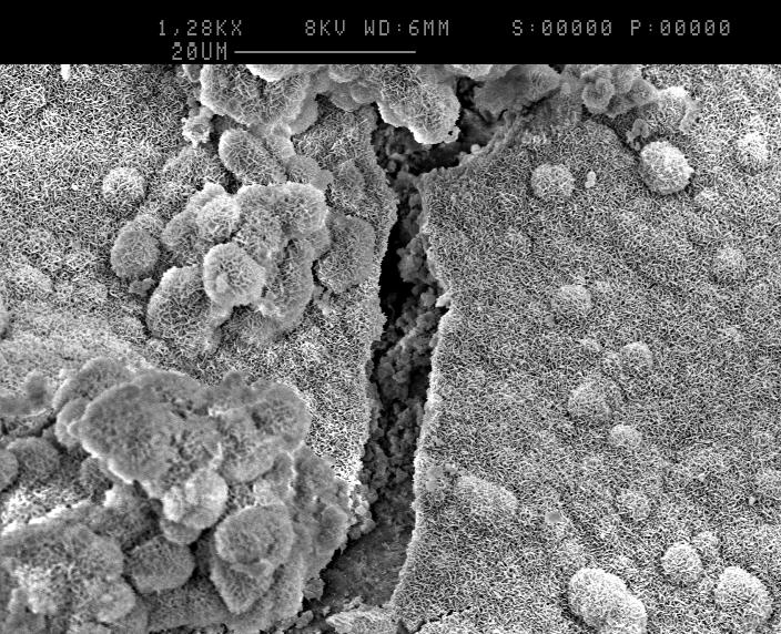
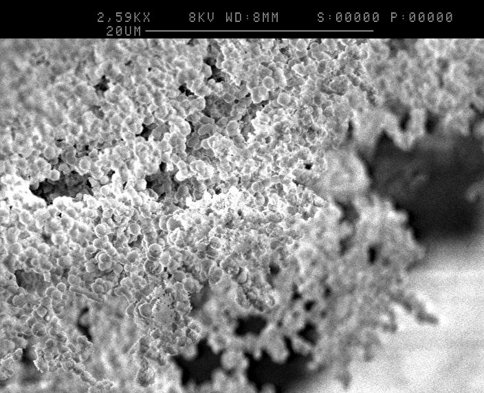
Scanning Electron Microscope Images

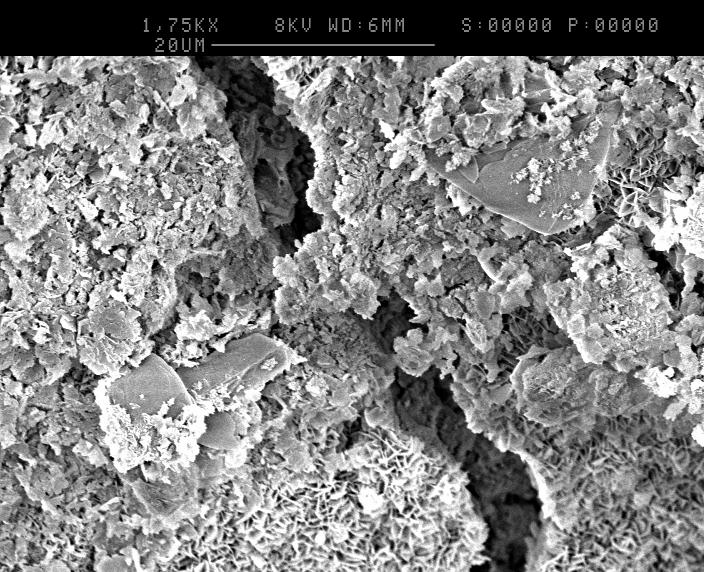
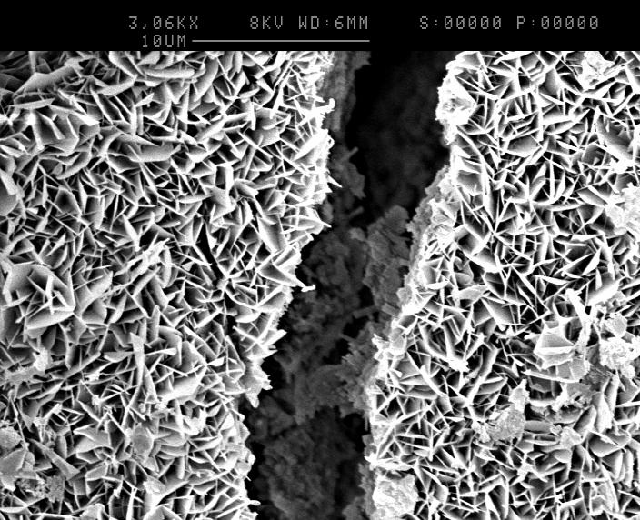
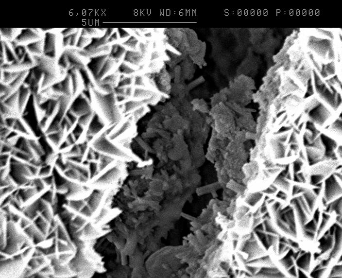
The Images below show Scanning electron microscope images of what our product should look like. Images 1-7 show an increasing magnification of a crack we are trying to fill.
Newcastle SEM4.jpgNewcastle SEM9.jpgNewcastle SEM6.jpg
(Images above: left to right)
  • Image1: shows our control concrete with no bacteria on the surface.
  • Image2: shows the surface of the concrete covered in calcium carbonate, glue and cells.
  • Image3: at this magnification you can begin to see calcium carbonate crystals filling a crack.
Newcastle SEM3.jpgNewcastle SEM8.jpgNewcastle SEM1.jpg
(Images above: left to right)
  • Image4: shows a higher magnification of calcium carbonates crystals and the crack which is being filled.
  • Image5: shows a crack almost completely covered in calcium carbonate, cells and glue.
  • Image6: shows a high magnification, the structure of the calcium carbonate crystals is more clear and some cells are visible in the crack.
Newcastle SEM2.jpgNewcastle SEM7.jpgNewcastle SEM5.jpg
(Images above: left to right)
  • Image7: this very high magnification shows cells and glue in the crack covered in calcium carbonate.
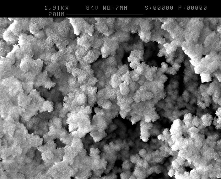
  • Image8: this image shows spores glued together with levan (induced by 10% sucrose)and calcium carbonate (produced by inducing with urea-calcium chloride broth). The cells have sporulated do to lack of nutrients and dehydration during preparation for SEM.
  • Image9: this images shows the spores are glued together as they are hanging from a vertical edge

An e-Science Approach to Synthetic Biology

We investigate the benefits of an e-Science approach, with a focus on workflows, to synthetic biology.