Team:Newcastle/23 July 2010
From 2010.igem.org

| |||||||||||||
| |||||||||||||
Contents |
Aims of genomic DNA extraction experiment
The aim of today's experiment is to extract genomic DNA from Bacillus subtilis strain 3610,genes from which will be needed for the swarming biobrick.
Materials Required
- Cells grown from yesterday
- Centrifuge
- pipette
- lysozyme
- Cell lysis solution
- RNase solution
- protein orecipitation solution
- ice
- isopropanol
- ethanol
Procedure
Cell lysis
- Pellet cells by centrifugation at 3600rpm for 10 minutes.
- Pour off supernatant.
- Add 0.5ml of cell suspension solution, gently pipet up and down to resuspend and transfer to 1.5 ml eppendorf tube.
- Add 25microlitres of lysozyme and invert 25 times.
- Incubate for 30 minutes at 37°C inverting occasionally.
- Centrifuge at 13000rpm for 10minutes to pellet the cells then remove the supernatant.
- Add 0.5ml of cell lysis solution to the cell pellet and gently pipet up and down to lyse the cells.
- Heat sample for 30 minutes mix every 5-10 minutes.
RNase treatment
- Add 3 microlitres of RNase A solution to the cell lysate
- Mix by inverting 25 times and incubate at 37°C for 60 minutes
Protein precipitation
- Cool samples on ice.
- Add 0.5 ml protein precipitation solution to each tube.
- Vortex vigorously at high speed for 20 seconds to miux the protein precipitation solution uniformly with the cell lysate. Place samples on ice for 5 minutes.
- Centrifuge at 13000 rpm for 30 seconds or until the precipitated proteins form a tight pellet.
DNA precipitation
- Pour the supernatant containing the DNA into a clean eppendorf tube. (The samples may be kept at -20°C overnight at this stage.)
- Add 0.5ml isopropanol to each tube.
- Mix by inverting gently for 50 times.
- Centrifuge at 13000 rpm for 1 minute. The DNA should be visible as a small white pellet.
- Pour off the supernatant and drain the tube on clean absorbent paper. Add 0.5 ml 70% ethanol and invert tube several times to wash the DNA.
- Centrifuge at 13000 rpm for 1 minute. Carefully pour off the ethanol.
- Drain the tubes on clean absorbent paper. Allow to air dry for 10-15 minutes.
DNA hydration
- Add 100 µl DNA hydration solution to each tube.
- Rehydrate DNA by incubating the sample for 1 hour at 65°C and overnight at room temperature. Tap the tube periodically to aid in dispersing the DNA.
- For storage, centrifuge briefly and store at -20°C.
Discussion
At the end of the DNA rpecipitation step, we did not observe any small white pellet.
Conclusion
If the experiment has failed the experiment will be redone on Monday, 26th July, 2010.
Aim of Arginine experiment
The aim of this experiment is to determine whether B. subtilis 168 is able to take up external arginine.
Procedure
- For the experimental protocol, please see 22.07.10 lab notebook.
Results
Arginine is an amino acid that is acidic. Therefore if B. subtilis 168 is able to take up arginine, it will cause a pH change in the media. This would result in an increase in pH.
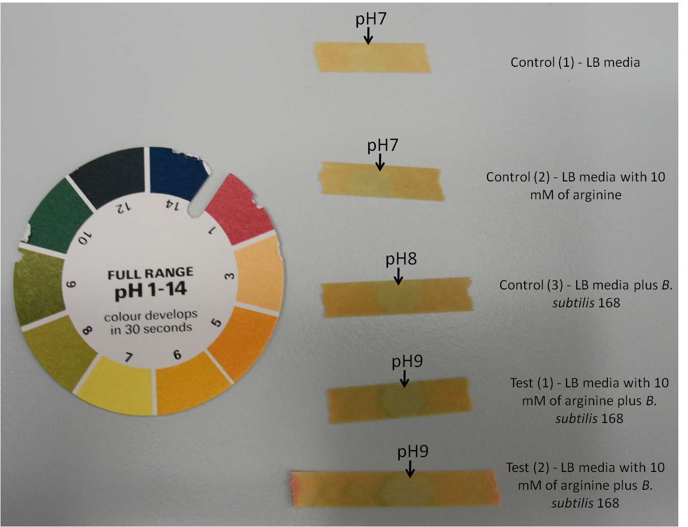
Figure 1: Arginine test using pH indicator stick to measure pH changes in the media.
| Time (in minutes) | Control (1) | Control (2) | Control (3) | Test (1) | Test (2) |
|---|---|---|---|---|---|
| 0 | pH 7 | pH 7 | pH 7 | pH 7 | pH 7 |
| 30 | pH 7 | pH 7 | pH 7 | pH 7 | pH 7 |
| 60 | pH 7 | pH 7 | pH 7 | pH 7 | pH 7 |
| 3 | pH 7 | pH 7.5 | |||
| 4 | pH 7 | pH 8 | |||
| 5 | pH 7 | pH 8 |
- Control (1) - LB media
- Control (2) - LB media with 10 mM of arginine
- Control (3) - LB media plus B. subtilis 168
- Test (1) - LB media with 10 mM of arginine plus B. subtilis 168
- Test (2) - LB media with 10 mM of arginine plus B. subtilis 168
At time point 0 min.
- Control (1) - pH 7
- Control (2) - pH 7
- Control (3) - pH 7
- Test (1) - pH 7
- Test (2) - pH 7
At time point 30 min.
- Control (1) - pH 7
- Control (2) - pH 7
- Control (3) - pH 7
- Test (1) - pH 7
- Test (2) - pH 7
At time point 60 min.
- Control (1) - pH 7
- Control (2) - pH 7
- Control (3) - pH 7
- Test (1) - pH 7
- Test (2) - pH 7
At time point 90 min.
- Control (1) - pH 7
- Control (2) - pH 7
- Control (3) - pH 7
- Test (1) - pH 7
- Test (2) - pH 7
At time point 120 min.
- Control (1) - pH 7
- Control (2) - pH 7
- Control (3) - pH 7
- Test (1) - pH 8
- Test (2) - pH 8
At time point 150 min.
- Control (1) - pH 7
- Control (2) - pH 7
- Control (3) - pH 7
- Test (1) - pH 8
- Test (2) - pH 8
At time point 180 min.
- Control (1) - pH 7
- Control (2) - pH 7
- Control (3) - pH 7
- Test (1) - pH 9
- Test (2) - pH 9
At time point 210 min.
- Control (1) - pH 7
- Control (2) - pH 7
- Control (3) - pH 7
- Test (1) - pH 9
- Test (2) - pH 9
At time point 240 min.
- Control (1) - pH 7
- Control (2) - pH 7
- Control (3) - pH 7
- Test (1) - pH 9
- Test (2) - pH 9
At time point 270 min.
- Control (1) - pH 7
- Control (2) - pH 7
- Control (3) - pH 8
- Test (1) - pH 9
- Test (2) - pH 9
At time point 300 min.
- Control (1) - pH 7
- Control (2) - pH 7
- Control (3) - pH 8
- Test (1) - pH 9
- Test (2) - pH 9
Conclusion
B. subtilis 168 breakdowns arginine to urea by producing arginase. The urea is then further broken down to ammonia and carbonate. This will lead to an increase in pH. Both the test 1 and test 2, which contain B. subtilis 168 and 10 mM of arginina show an increase in pH from 7 to 9. While the control 1 and control 2, which contain no B. subtilis 168 remains at pH 7. The control 3 which contain B. subtilis 168 but without addition of arginine show an increase in pH from 7 to 8. This could be due to unidentified products that are secreted by the bacteria.
Therefore this experiment have shown that B. subtilis 168 is able to utilise arginine, and thus increase the overall pH of the media. The overalll change in pH will cause calcium ions to precipitate with carbonate, forming calcium carbonate.
 
|
 "
"
