Team:Heidelberg/Notebook/MiMeasure/October

From 2010.igem.org

Revision as of 03:29, 28 October 2010 by Ting (Talk | contribs)

July
MTWTFSS
1234
567891011
12131415161718
19202122232425
262728293031

-


August
MTWTFSS
1
2345678
9101112131415
16171819202122
23242526272829
3031


September
MTWTFSS
12345
6789101112
13141516171819
20212223242526
2728293031

-


October
MTWTFSS
123
45 678910
11131415161718
19202122232425
2627282930

-


21/10/2010

seeding HeLa and Huh7 in 96-well plates

22/10/2010

Transfection

23/10/2010

seeding HeLa and Huh7 in 96-well plates

24/10/2010

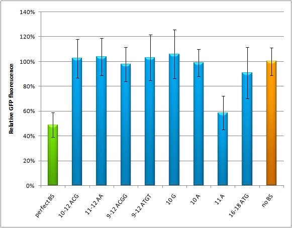
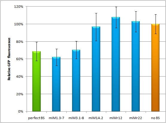
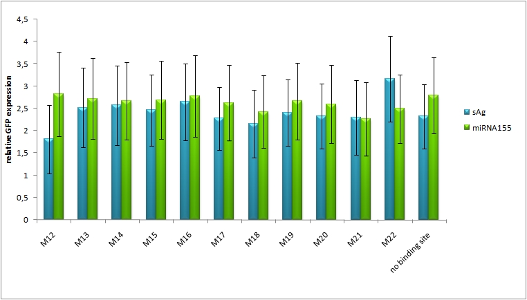
HeLa cells were transfected with miMeasure constructs with GFP expression regulated by different imperfect binding sites for miRsAg. As controls we used miMeasure construct with perfect binding site and no binding site. Each miMeasure was cotransfected with pcDNA plasmid expressing synthetic miRsAg and pcDNA5 expressing synthetic shRNA3.


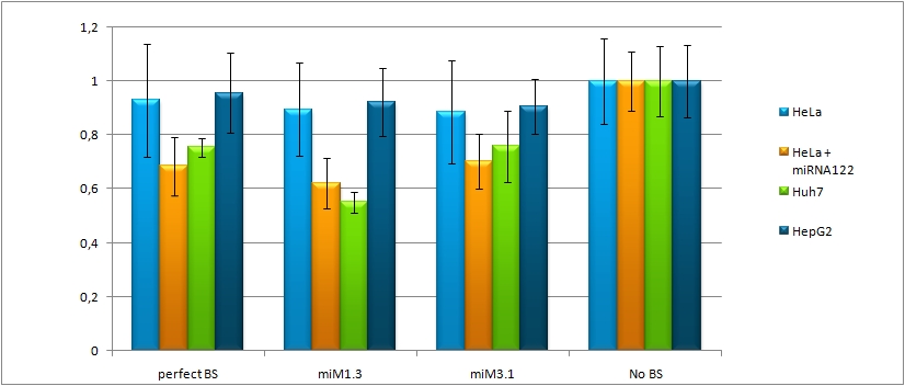
Transfection of HeLa and Huh7 cells with miMeasure with binding sites for miR122.

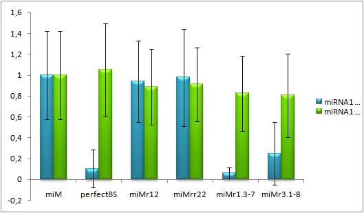
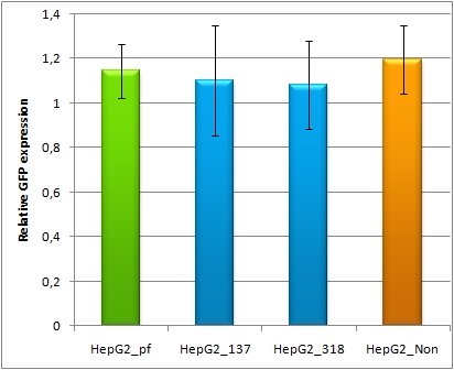
Huh7 were transfected with 50ng of miMeasure. HeLa cells were used as a control they were transfected with miMeasure constructs and miR122. Control without miR122 expression plasmid was made.

10/26/2010

microscopy measurements

different binding sites for miRsAg
different binding sites for miR122, HeLa cotransfected with miR122 expression plasmid
  • Flow cytometry measurements
    • HeLa and HuH7 cells were seeded two days. One day before the cells were transfected and 100µl of cells are used for the flow cytometry, whereby 10000 cells are measured for each sample.
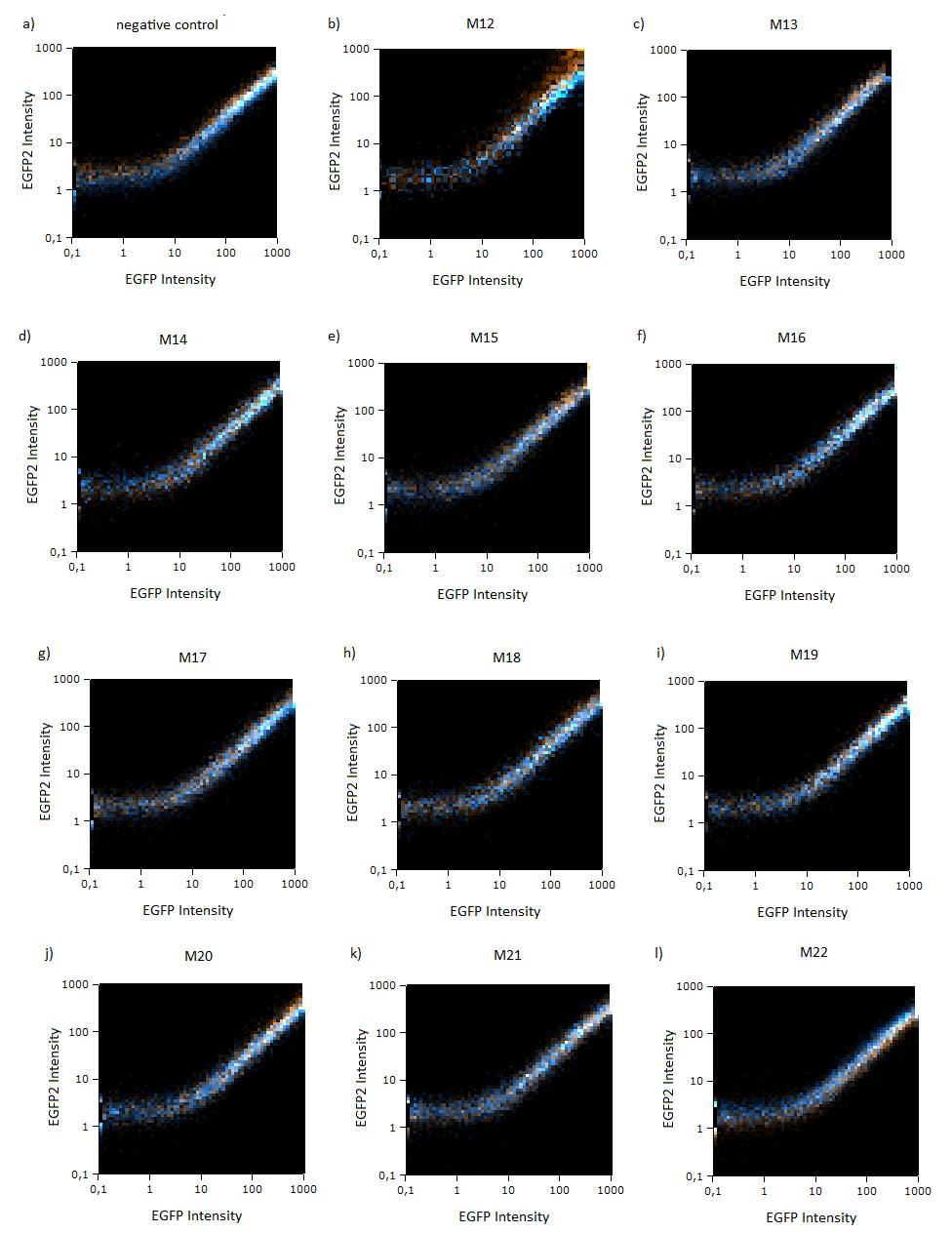
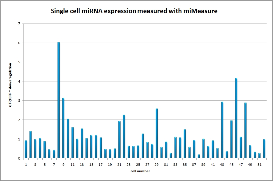
GFP/BFP ratio of single cells according to flow cytometry analysis
Cell lines mir122.jpg
Hela miRNA122 ratio.jpg
HepG2 miR122 microscopy.jpg
SAg miR155 HeLa.jpg
Single cellmiRNA expression.jpg
Single cellmiRNA expression cells.jpg

10/27/2010

HuH7 cells seeded and trasnfected one day before are imaged under the confocal microscope.

  • constructs:
    • perfect binding sites against miR-122
    • 3 perfect binding sites aligned via miRaPCR
    • 2 imperfect binding sites aligned via miRaPCR


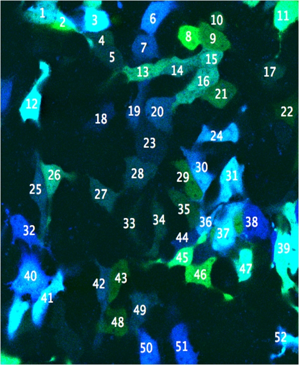
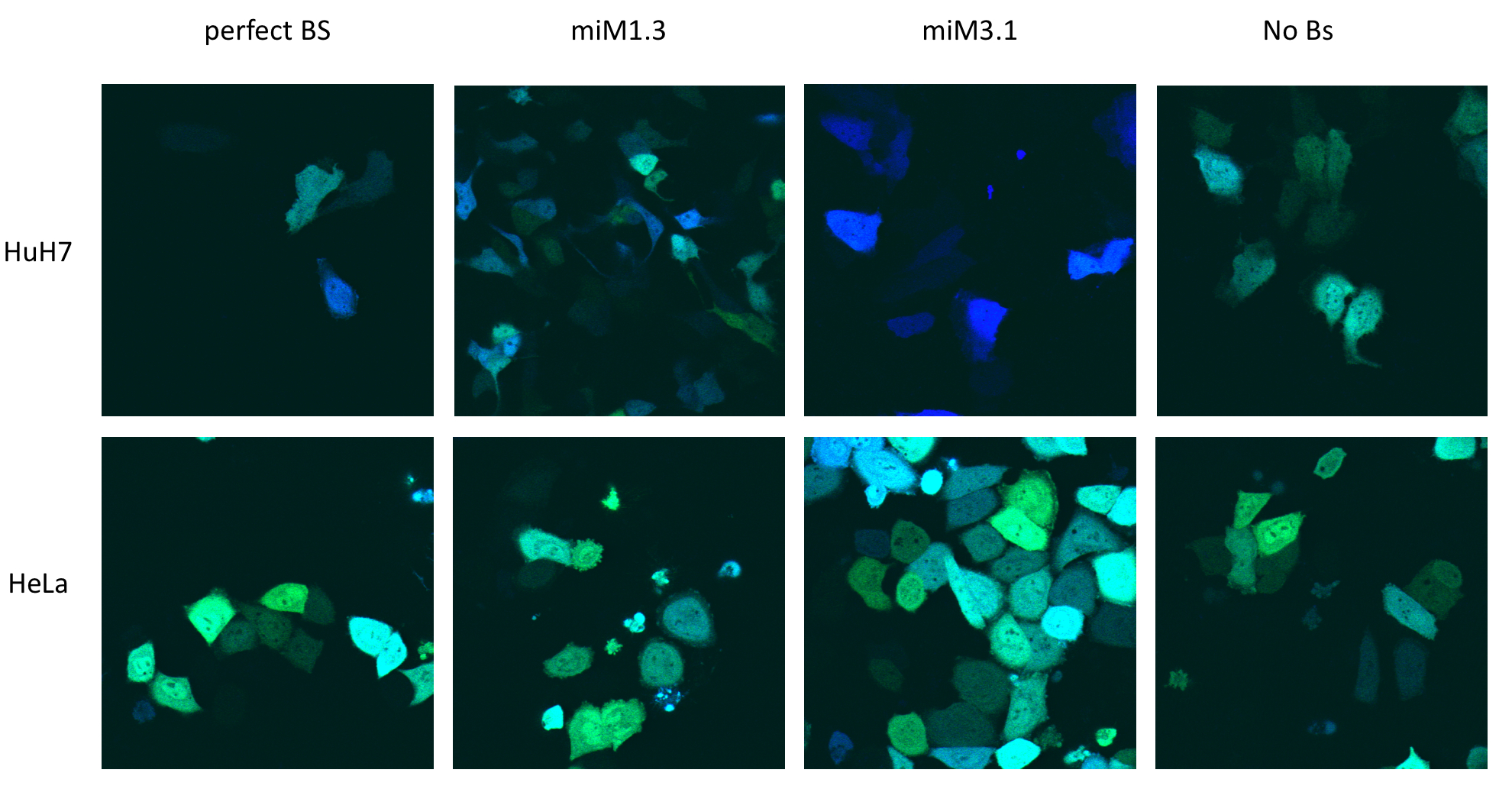
HuH7 cells transfected with different binding sites are imaged with the confocal microscope; 63x; green = EGFP, blue = EBFP2


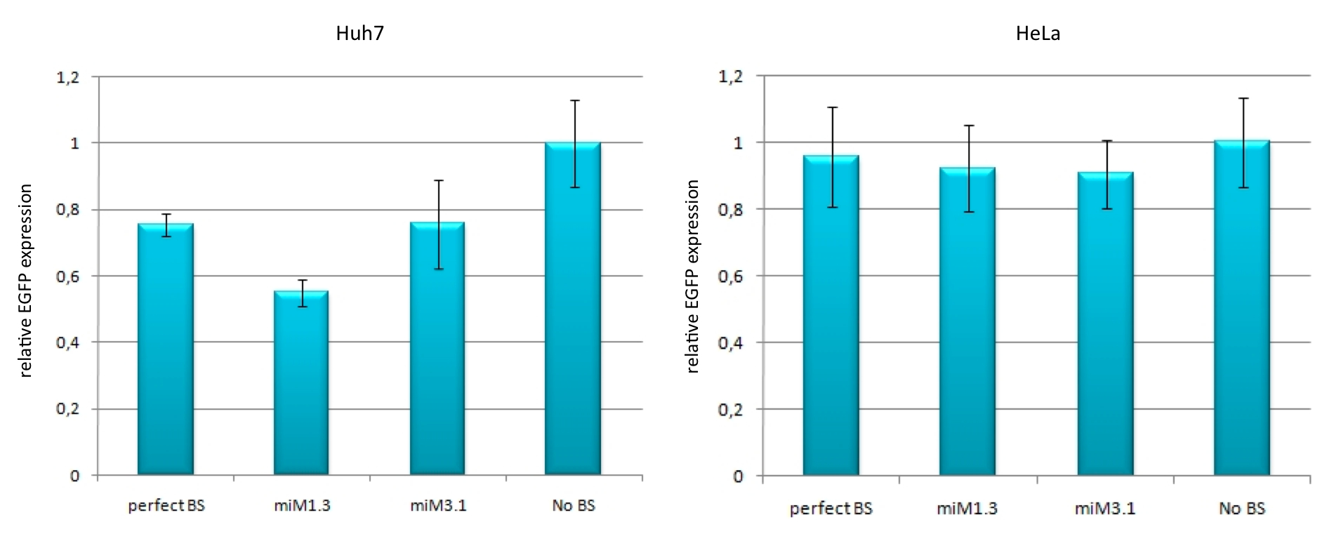
HeLa cells are also imaged under the confocal microscope and compared to the HuH7 cells in terms of EGFP/EGFP ratio normalized to the negative control.

comparison of HuH7 and HeLa cells": cells were transfected with the different constructs.