Team:Lethbridge/Modeling
From 2010.igem.org
Liszabruder (Talk | contribs) |
Liszabruder (Talk | contribs) |
||
| Line 125: | Line 125: | ||
=<font color="white">Homology Modeling= | =<font color="white">Homology Modeling= | ||
| - | |||
<html> | <html> | ||
<body> | <body> | ||
| Line 133: | Line 132: | ||
<tr> | <tr> | ||
<th> | <th> | ||
| - | <img src="https://static.igem.org/mediawiki/2010/ | + | <img src="https://static.igem.org/mediawiki/2010/6/6f/Homology1.png" width="300"/> |
</th> | </th> | ||
</tr> | </tr> | ||
| Line 154: | Line 153: | ||
[[Image:homology2.png|x350px|right|text-top]] | [[Image:homology2.png|x350px|right|text-top]] | ||
| + | <html> | ||
| + | <body> | ||
| + | <table border="0" cellpadding="8" width="28%" style="background-color:#000000" align="left"> | ||
| + | |||
| + | <tr> | ||
| + | <th> | ||
| + | <img src="https://static.igem.org/mediawiki/2010/6/6f/Homology1.png" width="300"/> | ||
| + | </th> | ||
| + | </tr> | ||
| + | |||
| + | </tr> | ||
| + | <tr><th colspan="1" align="left"> | ||
| + | <p> | ||
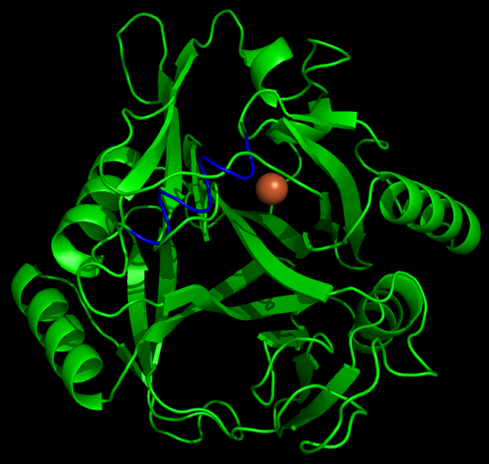
| + | <font color="white">Figure 2: Energy minimized position of the N-terminal 10X Arg tag (shown in blue). | ||
| + | </th> | ||
| + | </tr> | ||
| + | |||
| + | </table> | ||
| + | </body> | ||
| + | </html> | ||
To model the xylE structure, the sequence for xylE from <i>Pseudomonas putida</i> (NCBI accession number NP_542866) was aligned with the primary sequence from the crystal structure of xylE from the same organism (pdb: 1MPY; several differences in amino acid sequence were observed) using CLUSTALW (Higgins <i>et al.</i>, 1996). Based on this sequence alignment, a homology model was generated using the alignment mode in SWISSMODEL (Guex <i>et al.</i>, 1997; Kiefer <i>et al.</i>, 2009; Arnold <i>et al.</i>, 2006). To model the placement of an N-terminal arginine tag, the tag was manually added to the N-terminus of the model. Energy minimization was carried out in SWISS-PDB viewer in vacuo utilizing a GROMOS96 energy minimization (Guex <i>et al.</i>, 1997). The resulting pdb file was visualized and manipulated using PYMOL, images were taken using the same software (DeLano, 2006). | To model the xylE structure, the sequence for xylE from <i>Pseudomonas putida</i> (NCBI accession number NP_542866) was aligned with the primary sequence from the crystal structure of xylE from the same organism (pdb: 1MPY; several differences in amino acid sequence were observed) using CLUSTALW (Higgins <i>et al.</i>, 1996). Based on this sequence alignment, a homology model was generated using the alignment mode in SWISSMODEL (Guex <i>et al.</i>, 1997; Kiefer <i>et al.</i>, 2009; Arnold <i>et al.</i>, 2006). To model the placement of an N-terminal arginine tag, the tag was manually added to the N-terminus of the model. Energy minimization was carried out in SWISS-PDB viewer in vacuo utilizing a GROMOS96 energy minimization (Guex <i>et al.</i>, 1997). The resulting pdb file was visualized and manipulated using PYMOL, images were taken using the same software (DeLano, 2006). | ||
 "
"


