Project/Venus/
From 2010.igem.org
| Line 13: | Line 13: | ||
[[Image:Venus_digest.JPG]] <br /> <br /> | [[Image:Venus_digest.JPG]] <br /> <br /> | ||
| - | The Venus fragment was ligated into the linearised form of PSB1C3 (digested with EcoR1 and Pst1) and the re-digested with EcoR1 and Pst1 so to confirm the successful ligation. <br /> | + | The Venus fragment was ligated into the linearised form of PSB1C3 (digested with EcoR1 and Pst1) and the re-digested with EcoR1 and Pst1 so to confirm the successful ligation. The 782bp fragment confirms the ligation of Venus while the 2072bp fragment is indicative of the PSB1C3 backbone. <br /> |
[[Image:Venus_in_PSB1C3.JPG]] <br /> <br /> | [[Image:Venus_in_PSB1C3.JPG]] <br /> <br /> | ||
| Line 23: | Line 23: | ||
=== Testing and verification === | === Testing and verification === | ||
<br /> | <br /> | ||
| + | === References === | ||
| + | [1]Marco Weinberg, PhD. Antiviral Gene Therapy Research Unit, Department of Molecular Medicine and Haematology, University of the Witwatersrand Medical School, 7 York Rd. Parktown 2193 SOUTH AFRICA, marc.weinberg@wits.ac.za. | ||
Revision as of 10:43, 24 October 2010

Contents |
Characterisation of the fluorescent reporter protein - Venus - [http://partsregistry.org/Part:BBa_K354002 BBa_K354002]
BioBrick Background
Sequence Development and Annotation
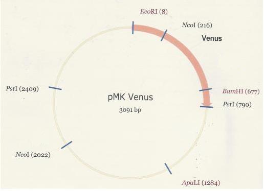
As was the case in the assembly of the PlcR-PapR BioBrick, the synthesized Venus BioBrick was received in the pMK plasmid (below).

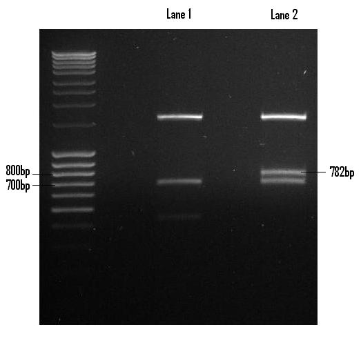
Once digested with EcoR1 and Pst1, the Venus fragment was recovered via gel extraction and purified in preparation for its ligation into PSB1C3. Lane 2 houses the Venus digest with the excised fragment shown (782bp).

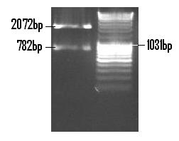
The Venus fragment was ligated into the linearised form of PSB1C3 (digested with EcoR1 and Pst1) and the re-digested with EcoR1 and Pst1 so to confirm the successful ligation. The 782bp fragment confirms the ligation of Venus while the 2072bp fragment is indicative of the PSB1C3 backbone.

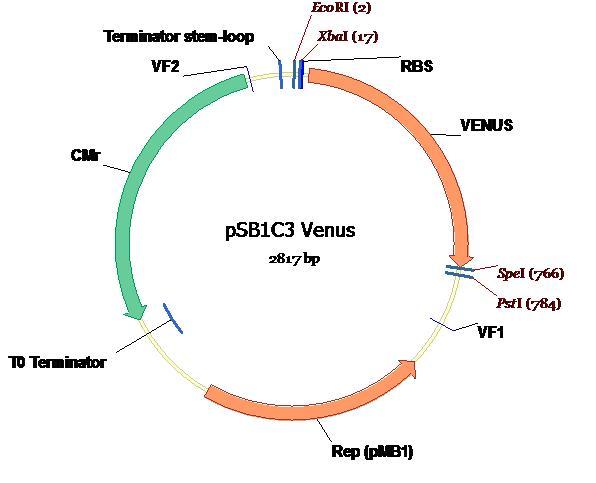
The completed ligation of Venus into the PSB1C3 plasmid is depicted on the plasmid map (below) along with all annotations and landmark features [1].

Testing and verification
References
[1]Marco Weinberg, PhD. Antiviral Gene Therapy Research Unit, Department of Molecular Medicine and Haematology, University of the Witwatersrand Medical School, 7 York Rd. Parktown 2193 SOUTH AFRICA, marc.weinberg@wits.ac.za.
 "
"