Team:HokkaidoU Japan/Notebook/September13
From 2010.igem.org
(Difference between revisions)
(→Gel purification of GFP(1-12O) and concentration check) |
|||
| (3 intermediate revisions not shown) | |||
| Line 10: | Line 10: | ||
<div style="clear:both;"></div> | <div style="clear:both;"></div> | ||
| - | =Electrophoresed after gel extraction= | + | =Electrophoresed after [[Team:HokkaidoU_Japan/Protocols|gel extraction]]= |
[[Image:HokkaidoU Japan 20100913b.jpg|200px|right|thumb|Electrophoresis after purification]] | [[Image:HokkaidoU Japan 20100913b.jpg|200px|right|thumb|Electrophoresis after purification]] | ||
| - | * Electrophoresed | + | * Electrophoresed [https://2010.igem.org/Image:HokkaidoU_Pictures_DNA_Marker.png TSUDA I] 2 uL and 0.5 uL of purified solution |
→Estimated concentration to be 54 ng/uL | →Estimated concentration to be 54 ng/uL | ||
* This time band location is good | * This time band location is good | ||
Latest revision as of 08:15, 27 October 2010
araC promoter purification
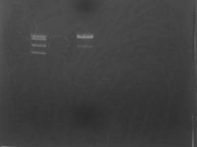
Compared to marker band was a little lower than it should. Thinking that this was due to too big an amount, gel extracted anyway.
- Used TSUDA marker
- Part length is 1259 bp
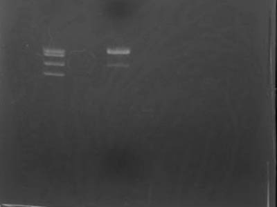
Electrophoresed after gel extraction
- Electrophoresed TSUDA I 2 uL and 0.5 uL of purified solution
→Estimated concentration to be 54 ng/uL
- This time band location is good
- Accidentally excised a part of other band resulting small contamination
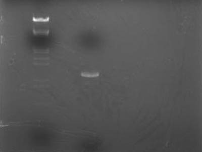
Gel purification of GFP(1-12O) and concentration check
- TSUDA I 2 uL, DNA 0.5 uL
→Estimated concentration to be 120 ng/uL
- Part length is 878 + 220 bp = 947 bp
- That slightly above mark so OK.
 "
"


