Team:HokkaidoU Japan/Notebook/August26
From 2010.igem.org
(Difference between revisions)
(→50 ug/uLに濃縮したpSB1C3の電気泳動) |
|||
| (3 intermediate revisions not shown) | |||
| Line 2: | Line 2: | ||
<div class="linkbar"><div class="prev">[[Team:HokkaidoU_Japan/Notebook/August25|August 25]]</div>[[Team:HokkaidoU_Japan/Notebook|Notebook]]<div class="next">[[Team:HokkaidoU_Japan/Notebook/August27|August 27]]</div></div> | <div class="linkbar"><div class="prev">[[Team:HokkaidoU_Japan/Notebook/August25|August 25]]</div>[[Team:HokkaidoU_Japan/Notebook|Notebook]]<div class="next">[[Team:HokkaidoU_Japan/Notebook/August27|August 27]]</div></div> | ||
| - | =50 ug/ | + | =Electrophoresis of pSB1C3 concentrated to 50 ug/uL= |
| - | [[Image:HokkaidoU Japan 20100826a.jpg|200px|right|thumb|]] | + | |
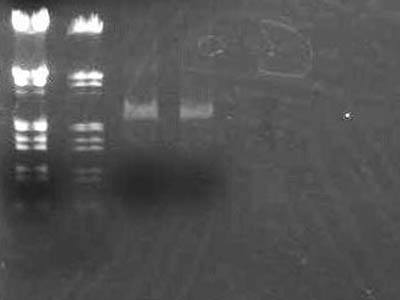
| - | * | + | [[Image:HokkaidoU Japan 20100826a.jpg|200px|right|thumb|Electrophoresis of concentrated pSB1C3]] |
| + | |||
| + | * Added 2.8 uL of 6x SB to 17.4 uL of pSB1C3 solution digested yesterday and electrophoresed | ||
| + | |||
{| class="protocol" | {| class="protocol" | ||
|- | |- | ||
| Line 11: | Line 14: | ||
|- | |- | ||
|1 | |1 | ||
| - | | | + | |Added too much of marker, mistake |
|- | |- | ||
|2 | |2 | ||
| - | |λ/''Hin''d III & EcoR I | + | |[https://2010.igem.org/Image:HokkaidoU_Pictures_DNA_Marker.png λ/''Hin''d III & EcoR I] |
|- | |- | ||
|3 | |3 | ||
| Line 23: | Line 26: | ||
|} | |} | ||
| - | * | + | * IF digestion and ligation went well there should be bands of dimers, trimers but none of the were visible |
| - | * | + | * Only band visible was monomer(about 2000 bp) |
| + | |||
| + | =Filtration of pSB1A3, pSB1C3 and pSB1K3 PCR solutions= | ||
| + | |||
| - | + | Remaining amount from check via electrophoresis,namely 49 uL was filtrated with Microcon YM-10 | |
| - | + | ||
Latest revision as of 07:54, 27 October 2010
Electrophoresis of pSB1C3 concentrated to 50 ug/uL
- Added 2.8 uL of 6x SB to 17.4 uL of pSB1C3 solution digested yesterday and electrophoresed
| Lane | DNA |
| 1 | Added too much of marker, mistake |
| 2 | λ/Hind III & EcoR I |
| 3 | pSB1C3 solution |
| 4 | pSB1C3 solution |
- IF digestion and ligation went well there should be bands of dimers, trimers but none of the were visible
- Only band visible was monomer(about 2000 bp)
 "
"


