Team:HokkaidoU Japan/Notebook/August13
From 2010.igem.org
(Difference between revisions)
| Line 29: | Line 29: | ||
|style="text-align:left;"| 1 uL | |style="text-align:left;"| 1 uL | ||
|- | |- | ||
| - | |style="text-align:right;"| Template ([[Team:HokkaidoU_Japan/ | + | |style="text-align:right;"| Template ([[Team:HokkaidoU_Japan/Materials_And_Methods#BioBricks|1-18F]]) : |
|style="text-align:left;"| 1 uL | |style="text-align:left;"| 1 uL | ||
|- | |- | ||
Revision as of 11:26, 16 October 2010
PCR of parts which didn't amplify well via mini prep
Composition of Reaction Solution
| Reagent | Amount |
|---|---|
| Autoclaved DW : | 33 uL |
| 10x PCR buffer : | 5 uL |
| 2 mM dNTPs : | 5 uL |
| 25 mM MgSO4 : | 3 uL |
| EX-F primer : | 1 uL |
| PS-R primer : | 1 uL |
| KOD plus Neo : | 1 uL |
| Template (1-18F) : | 1 uL |
| Total : | 50 uL |
Removal of Primers
- Added 150 uL of TE to 50 uL of PCR product, each
- Transfered into Microcon YM-10 filter and cetrifuged for 20 min at 14,000 G
- Much of the solution remained so centrifuged for aditional 10 min
- Measured the amount left
- It was 140 uL so centrifuged again for 10min
- And again
- Finally DNA solution was reduced to 19 uL so we added 31 uL to make final volume of 50 uL
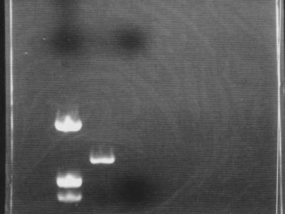
Electrophoresis
- Added 0.4 uL of 6x Sample Buffer to 1 uL of DNA solution and electrophoresed it.
- At the same time add added 2 uL of 6x Sample Buffer to 10 uL of flow trough and centrifuged
- Used marker pUC119/Hinf
- It was confirmed that DNA was amplified by PCR
- Between marker bands of 543 bp and 1330 bp PCR product band of 700 bp was visible
- Due to negligence (electrophoresis for 45 min), flow through with primers flowed through and exited gel
 "
"


