Team:Imperial College London/Modelling

From 2010.igem.org

(Difference between revisions)
m (Adding sentence)
m
Line 118: Line 118:
<b style="font-size:12px">Surface Protein Model</b><br/>
Goals:
Goals:
-
<ol><p>The aim of this model is to determine the concentration of Schistosoma elastase or TEV protease that should be added to bacteria to trigger the response.</p>
+
<ol><p>The aim of this model is to determine the concentration of Schistosoma elastase or TEV protease that should be added to bacteria to trigger the response. That was supposed to allow us to correlate required concentration for the activation with the concentration of Schistosoma elastase in the lake.<</p>
<p>It is also attempted to model how long it takes for the protease or elastase to cleave enough peptides.</p>  
</ol>
</ol>

Revision as of 21:01, 13 October 2010

Temporary sub-menu:

Dry Lab Diary; Surface Protein Model; Output Amplification Model;Wet-Dry Lab Interaction

Introduction to modelling
In the process of designing our construct two major questions arose which could be answered by computer modelling:
  1. Output Amplification Model
    We came up with an idea of using amplification of colour output to make it show within minutes after stimulus. The question that arose concerned whether amplification will actually perform better than simple production in the cellular environment. Furthermore, we had trouble deciding whether we should design the amplification module to be consisting of 1,2 or even more amplification steps. It appeared that the problem was recognised to be difficult enough to employ modelling.
  2. Surface Protein Model
    We came up with novel idea of detecting organisms that we do not have a specific receptor for. In the particular example that we have been considering, Schistosoma's protease was meant to cleave designed by us protein displayed on bacteria's cell wall. The cleaved peptide was supposed to be recognised by the receptor which would act to activate the colour expression. That solution raised questions about the risk of false positive or whether in there are any chances for ComD receptors to get activated in the diluted environment. Modelling was recognised as suitable to answer those questions.
Results & Conclusions
Output Amplification Model
  1. It was concluded that there is no advantage of 2 step amplification over 1 step amplification in given context. With that conclusion design of 2-step amplifier was abandoned.
  2. It could not be firmly decided whether 1-step amplification is going to speed up colour expression over simple production or not. It is because it starts off slower but then sharply rises to result in higher active dioxygenase concentrations.
  3. Conditions for amplification to be effective were determined.
Graph showing advantage of 1 ammplification output over simple production
Concentration of coloured compound.


Surface Protein Model

  1. Initial TEV protease concentrations we determined for the optimal activation of the receptor within 1.5 minute after elastases would have come into contact with our cell.
Graph showing when threshold AIP concentration is reached (for different initial TEV concentrations). Notice log-log scale.
Graph showing when threshold AIP concentration is reached
(for different initial TEV concentrations). Notice log-log scale.
Quick overview of models
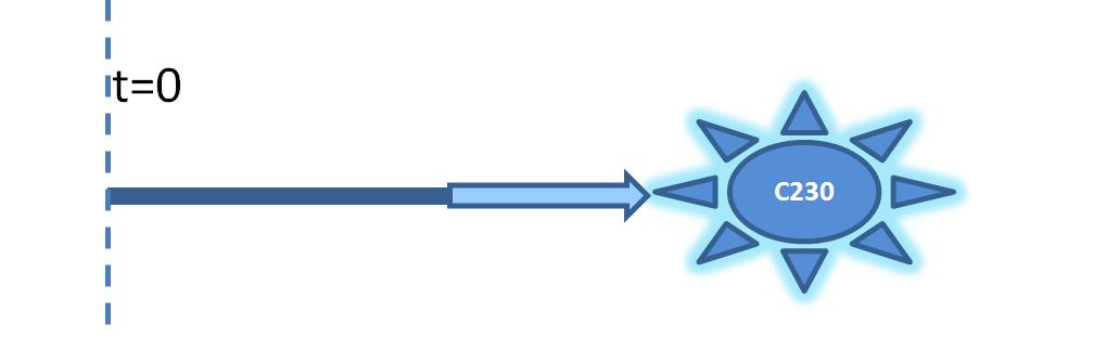
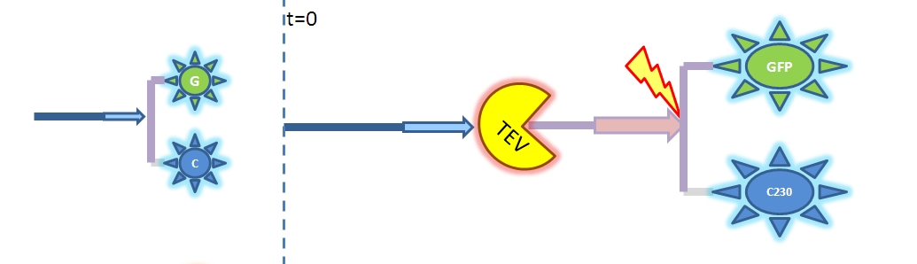
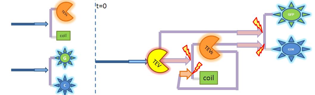
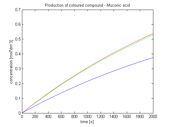
Output Amplification Model

Goals:

    This model was mainly developed in order to determine whether simple production is better than 1- or 2-step amplification.

    Further goals, contained estimation of the speed of modelled response.

Elements of the system:

  1. Dioxygenase (blue on diagrams below) is an enzyme that acts on catechol to produce yellow output. In most of our models dioxygenase was treated as an output, actually, as it was found that active dioxygenase acting on catechol produces yellow colour within split second.
  2. GFP-Dioxygenase fusion protein (GFP is shown green on diagrams). Dioxygenase joined by the linker to GFP was assumed to be inactive.
  3. TEV protease (yellow on diagrams below) has ability to cleave GFP-Dioxygenase fusion protein, hence, activate the dioxygenase
  4. Split TEV protease (orange on diagrams below), is an inactive, split form of TEV mounted on coiled coils. It can be activated again by coiled coils being cleaved by another active TEV.


Diagram of simple production of Dioxygenase
Diagram illustrating simple production of dioxygenase.
Diagram of simple production of Dioxygenase
Diagram showing 1 step amplification. TEV enzyme is being simply produced to activate pre-produced inactive dioxygenase.
Diagram illustrating 2 step amplification. Enzyme is activating enzyme which activates dioxygenase. Both pre-products have same TEV-site, so simply produced TEV is allowed to act directly on dioxygenase too.


Major assumptions:

  1. The chemical and enzymatic reactions are modelled according to the Law of Mass Action.
  2. Model assumes that the modelled system is innert within bacterial body or that reaction with outher species within bacterium is neglible. For example, TEV protease is supposed to be unsuccessful in cleaving other molecules due to its specifity.


Surface Protein Model
Goals:

    The aim of this model is to determine the concentration of Schistosoma elastase or TEV protease that should be added to bacteria to trigger the response. That was supposed to allow us to correlate required concentration for the activation with the concentration of Schistosoma elastase in the lake.<

    It is also attempted to model how long it takes for the protease or elastase to cleave enough peptides.

Elements of the system:

  1. Surface protein that consists of cell wall binding domain, linker, AIP (Auto Inducing Peptide)
  2. Schistosoma elastase (enzyme released by the parasite) cleaves AIP from cell wall binding domain at the linker site. In laboratory we used TEV protease as we could not get handle of Schistosoma elastase.
  3. ComD receptor is being activated by high enough AIP concentration.

Major assumptions:

  1. The chemical and enzymatic reactions are modelled according to the Law of Mass Action.
  2. Model assumes that the modelled system is innert within bacterial body or that reaction with outher species within bacterium is neglible. For example, TEV protease is supposed to be unsuccessful in cleaving other molecules due to its specifity.
  3. Due to carefully chosen cell concentrations, the diffusion of free AIPs could be neglected. However, that restricts the model to the considered cell concentrations only.
  4. Receptor activation threshold was defined by 1 specific value as opposed to considering intermediate states between fully "off" and "on".