 "
"
File:Homology1.png
From 2010.igem.org
(Difference between revisions)
David.franz (Talk | contribs) (uploaded a new version of "Image:Homology1.png") |
David.franz (Talk | contribs) (uploaded a new version of "Image:Homology1.png") |
Revision as of 18:22, 23 October 2010
Homology model of XylE from Pseudomonas putida. The original structure (1MPY) is shown in teal; the generated homology model is shown in green. An Fe2+ ion near the proposed active site is shown is brown.
File history
Click on a date/time to view the file as it appeared at that time.
| Date/Time | Thumbnail | Dimensions | User | Comment | |
|---|---|---|---|---|---|
| current | 23:45, 24 October 2010 | 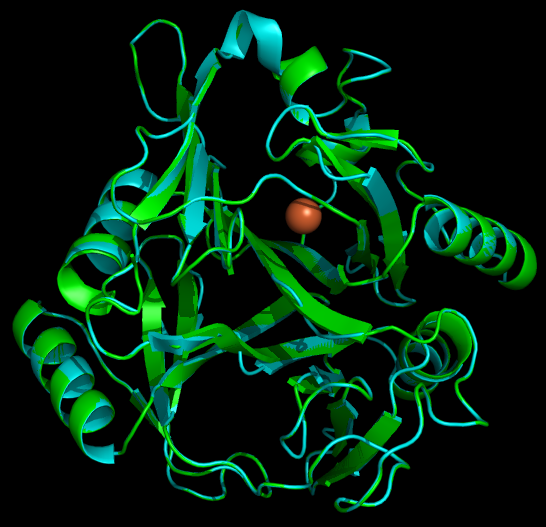 | 546×527 (152 KB) | Liszabruder (Talk | contribs) | |
| 18:27, 23 October 2010 |  | 807×819 (336 KB) | David.franz (Talk | contribs) | ||
| 18:24, 23 October 2010 |  | 845×797 (334 KB) | David.franz (Talk | contribs) | ||
| 18:22, 23 October 2010 |  | 848×852 (298 KB) | David.franz (Talk | contribs) | ||
| 17:50, 23 October 2010 |  | 545×521 (140 KB) | David.franz (Talk | contribs) | ||
| 17:47, 23 October 2010 |  | 1,248×626 (161 KB) | David.franz (Talk | contribs) | (Homology model of XylE from Pseudomonas putida. The original structure (1MPY) is shown in teal; the generated homology model is shown in green. An Fe2+ ion near the proposed active site is shown is brown.) |
File links
There are no pages that link to this file.