Team:Stockholm/20 September 2010
From 2010.igem.org
Contents |
Andreas
Assembly of new parts
- pSB1K3.N-LMWP⋅SOD⋅His
- Dig pSB1C3.N-LMWP (E+A)
- Dig pMA.SOD⋅His (N+P)
- Dig pSB1K3.RFP (E+P)
- pSB1C3.N-LMWP⋅SOD⋅His
- Dig pSB1C3.N-LMWP (A+S)
- Dig pMA.SOD⋅His (N+S)
Digestions
| pSB1C3. N-LMWP | pMA. SOD⋅His | pSB1C3. N-LMWP | pSB1K3. N-TAT⋅SOD⋅ His 4 | |
|---|---|---|---|---|
| 10X FastDigest buffer | 3 | 3 | 3 | 2 |
| dH2O | 15.2 | 4.1 | 15.2 | 11.4 |
| DNA (1 μg) | 9.8 | 20.9 | 9.8 | 4.6 |
| AgeI | 1 | 0 | 1 | 0 |
| NgoMIV | 0 | 1 | 0 | 0 |
| FD SpeI | 1 | 1 | 0 | 0 |
| FD EcoRI | 0 | 0 | 1 | 0 |
| FD PstI | 0 | 0 | 0 | 1 |
| FD XbaI | 0 | 0 | 0 | 1 |
| 30 μl | 30 μl | 30 μl | 20 μl |
- Incubation: 37 °C, 2:00 (NgoMIV & AgeI); 0:30 (FD)
- Inactivation: 80 °C, 20 min
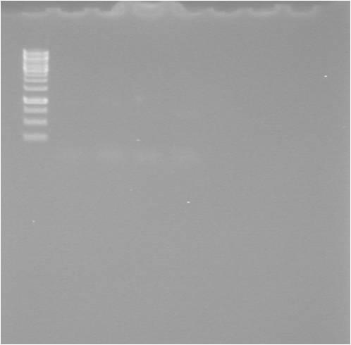
Gel verification
1.5 % agarose, 120 V
Expected bands
- Dig pSB1C3.N-LMWP A+S 20/9: 2118 bp, (14 bp)
- Dig pMA.SOD⋅His N+S 20/9: 2416 bp, 503 bp
- Dig pSB1C3.N-LMWP E+A 20/9: 2063 bp, 69 bp
- Dig pSB1K3.N-TAT⋅SOD⋅His 4 X+P 20/9: ≈2200 bp, 558 bp
Results
Ligations
- [Dig pSB1K3.RFP E+P 14/9] = 66.6 ng/μl
- [Dig pMA.His⋅SOD E+A 14/9] = 66.6 ng/μl
- [Dig pSB1C3.C-TAT N+P 15/9] = 66.6 ng/μl
- [Dig pSB1C3.N-LMWP A+S 20/9] = 33.3 ng/μl
- [Dig pMA.SOD⋅His N+S 20/9] = 33.3 ng/μl
- [Dig pSB1C3.N-LMWP E+A 20/9] = 33.3 ng/μl
- [Dig pSB1K3.N-TAT⋅SOD⋅His 4 X+P 20/9] = 33.3 ng/μl
| pSB1C3. N-LMWP⋅SH | pSB1K3. N-LMWP⋅SH | pSB1A2. RBS.yCCS | pEX. N-TAT⋅SH | |
|---|---|---|---|---|
| 10X T4 Ligase buffer | 2 | 2 | 2 | 2 |
| dH2O | 9 | 0 | 11 | 11 |
| Vector DNA | 2 | 1.5 | 1.5 | 1.5 |
| Insert 1 DNA | 6 | 4.5 | 4.5 | 4.5 |
| Insert 2 DNA | – | 11 | – | – |
| T4 DNA ligase | 1 | 1 | 1 | 1 |
| 20 μl | 20 μl | 20 μl | 20 μl |
- Incubation: 22 °C, 15 min
Transformations
Standard transformations, procedures according to protocol.
- 1 μl ligation mix
- Lig pSB1C3.N-LMWP⋅SH (Cm 25)
- Lig pSB1K3.N-LMWP⋅SH (Km 50)
- Lig pSB1A2.RBS.yCCS (Amp 100)
- Lig pEX.N-TAT⋅SH (Amp 100 + 50 μl 0.1 mM IPTG)
Mimmi
his.SOD.cTAT
Gel
| well | sample |
|---|---|
| 1 | ladder |
| 2 | pSB1C3.his.SOD.cTAT 1 |
| 3 | pSB1C3.his.SOD.cTAT 2 |
| 4 | pSB1C3.his.SOD.cTAT 3 |
| 5 | pSB1C3.his.SOD.cTAT 4 |
| 6 | pSB1C3.his.SOD.cTAT 5 |
| 7 | pSB1C3.his.SOD.cTAT 6 |
| 8 | pSB1C3.his.SOD |
| 9 | pEX.SOD |
PhastGel
| well | sample | |
|---|---|---|
| 1 | SOD.his 0h | |
| 2 | SOD.his 2h 1:1.5 | |
| 3 | his.SOD 0h | |
| 4 | his.SOD 2h 1:2 | |
| 5 | yCCS 1 2h 1:2 | |
| 6 | ladder |
pEX.SOD.his
Gel
| well | sample |
|---|---|
| 1 | ladder |
| 2 | pEX.SOD.his |
| 3 | pEX.his.SOD |
| 4 | yCCS 1 |
| 5 | yCCS 2 |
- Very weak bands, and in the wrong size...
Johan
- Cut tyrosinase with NgoMIV & SpeI
10 µl DNA
2 µl 10x buffer
1 µl NgoMIV
1 µl SpeI
6 µl H2O
- Cut bFGF with BamHI
2 µl DNA
2 µl 10x buffer
(1 µl BamHI)
15 µl H2O
Did a gel and showed that the bFGF are correct
- Ligated tyrosinase into pMA (vector with histag)
1 µl pMA
2 µl tyrosinase
2 µl 10x buffer
1 µl T4 ligase
14 µl H2O
 |

|
 |

|
 |

|

|

|
 "
"


