Team:EPF Lausanne/Project immuno
From 2010.igem.org


Contents |
Proteins
We have chosen two different ways to target the parasite P.falciparum and prevent the malaria transmission through mosquitos. Our engineered bacteria could express either an immunotoxin, or two p-proteins, or even both for maximum efficiency. We tried to express all of these proteins using the C3 plasmid incorporating the strong promoter, a constitutive sequence for greater level of expression.
The Immunotoxin is one of our tools to block transmission of malaria parasite in mosquitos. It is composed of two main parts : The first one is a single-chain antibody fragment (scFv) directed to Pbs2l, which is a surface membrane protein of Plasmodium berghei . The second part is a lytic peptide, Shiva-1, which acts by forming “pores” on the parasite’s membrane. The immunotoxin is supposed to specifically target and lyse the parasite.
In parallel to the immunotoxin we thought of a different way of blocking the P.falciparum by using a group of proteins, called the "p-proteins".
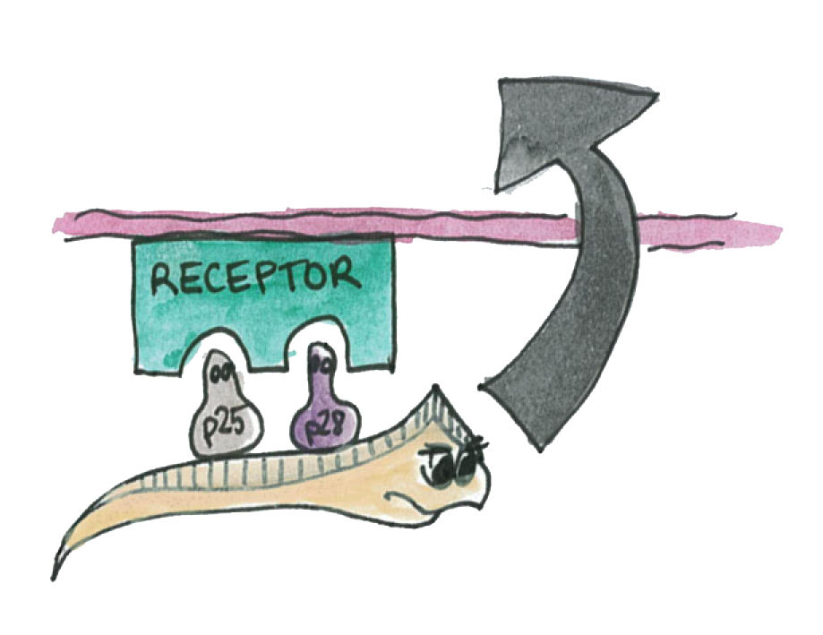
P25 and P28 are a class of important proteins expressed on the membrane of different type of Plasmodium; we call this ensemble of evolutionary conserved proteins the P-proteins. They are mainly expressed on the mosquito-stage parasite (ookinete).
The Plasmodium normally uses its P-protein to interact with the epithelium in order to go through it.
Now if Asaia starts to produce a high amout of these proteins, the interactions will be disrupted and the plasmodium will not propagate through the mosquito's epithelium.
Results
A: The Immunotoxin is expressed and appears in the supernatant
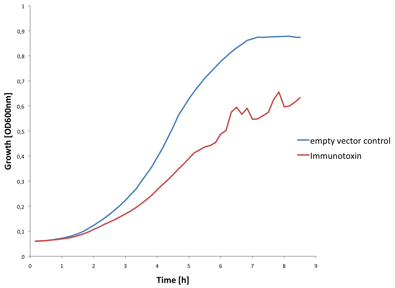
We tested expression of the immunotoxin in E. coli (see Materials and Methods for details). In a western blot analysis of whole cell lysates we could see bands corresponding to full length immunotoxin and possibly degraded fragments of the protein (see figure). The immunotoxin contains a PelB sequence that targets it for secretion into the periplasm. We concentrated the supernatant of both the immunotoxin and a control culture by running it through a filtering device with a 5 kDa cut-off. Running a western blot with these samples (see figure) we verified that the immunotoxin was also found in the supernatant.

B: The P-proteins are not expressed
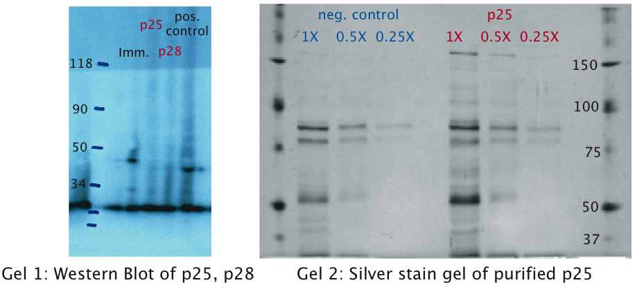
The same experiments were conducted for the p-proteins p25 and p28. No bands were detected on the western blots (see figure) which leads us to the conclusion that these proteins were only very weakly expressed or not at all. This might be explained by the fact that we took the native sequence from Plasmodium falciparum . The genome of Plasmodium falciparum is very A-T-rich ([http://areslab.ucsc.edu/cgi-bin/hgGateway UCSC Malaria Genome Browser]). We think that expression of p25 and p28 may be improved by codon optimizing it for expression in bacteria like E. coli and Asaia as we did with the immunotoxin. Additional to the Western Blots, to rule out the possibility that concentration was too low, we attempted a protein purification (following the [http://openwetware.org/wiki/Knight:Purification_of_His-tagged_proteins/Denaturing Knight protocol]) from a large culture volume (see figure) using Ni-NTA columns.
References
- [http://www.sciencedirect.com/science?_ob=ArticleURL&_udi=B6T29-42JHDJD-9&_user=10&_coverDate=03%2F31%2F2001&_rdoc=1&_fmt=high&_orig=search&_origin=search&_sort=d&_docanchor=&view=c&_acct=C000050221&_version=1&_urlVersion=0&_userid=10&md5=5f4b78b08a1846e241faaed33bc76cb3&searchtype=a 1. Shigeto Yoshida, Bacteria expressing single-chain immunotoxin inhibit malaria parasite development in mosquitoes, Molecular and Biochemical Parasitology (2001)]
- [http://www.nature.com/emboj/journal/v20/n15/full/7593895a.html 2. Ana M. Tomas, Gabriele Margos, Robert E. Sinden, P25 and P28 proteins of the malaria ookinete surface have multiple and partially redundant functions, The EMBO Journal (2001)]
- [http://www.ncbi.nlm.nih.gov/pmc/articles/PMC1951121/?tool=pubmed 3. Ajay K. Saxena, Yimin Wu, and David N. Garboczi, Plasmodium P25 and P28 Surface Proteins: Potential Transmission-Blocking Vaccines, Eukaryot Cell (2007)]
- [http://www.nature.com/nsmb/journal/v13/n1/full/nsmb1024.html 4. Ajay et al., The essential mosquito-stage P25 and P28 proteins from Plasmodium form tile-like triangular prisms, natures structural & molecular biology, 2005]

 "
"


