Team:Groningen/2 August 2010
From 2010.igem.org
Week 31
Induction of chaplins on agar plates
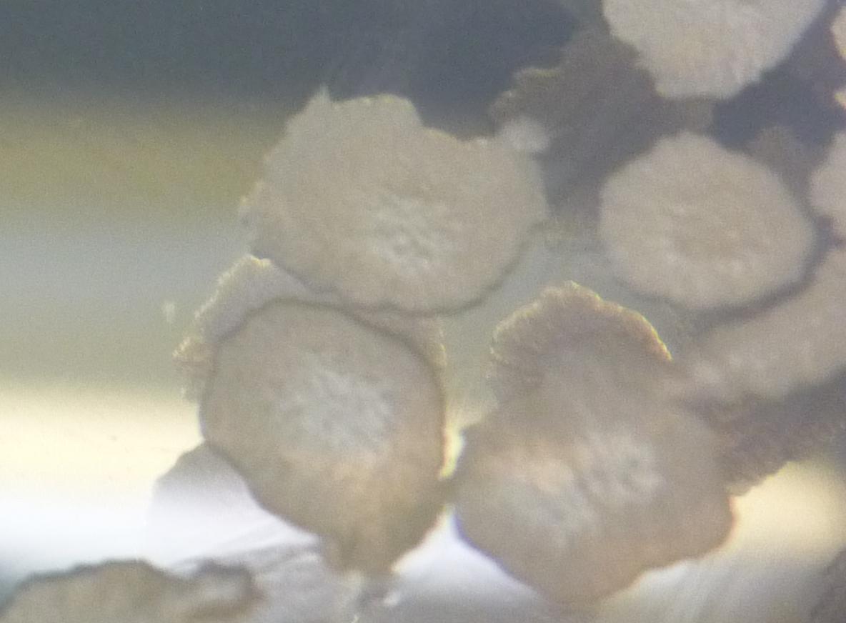
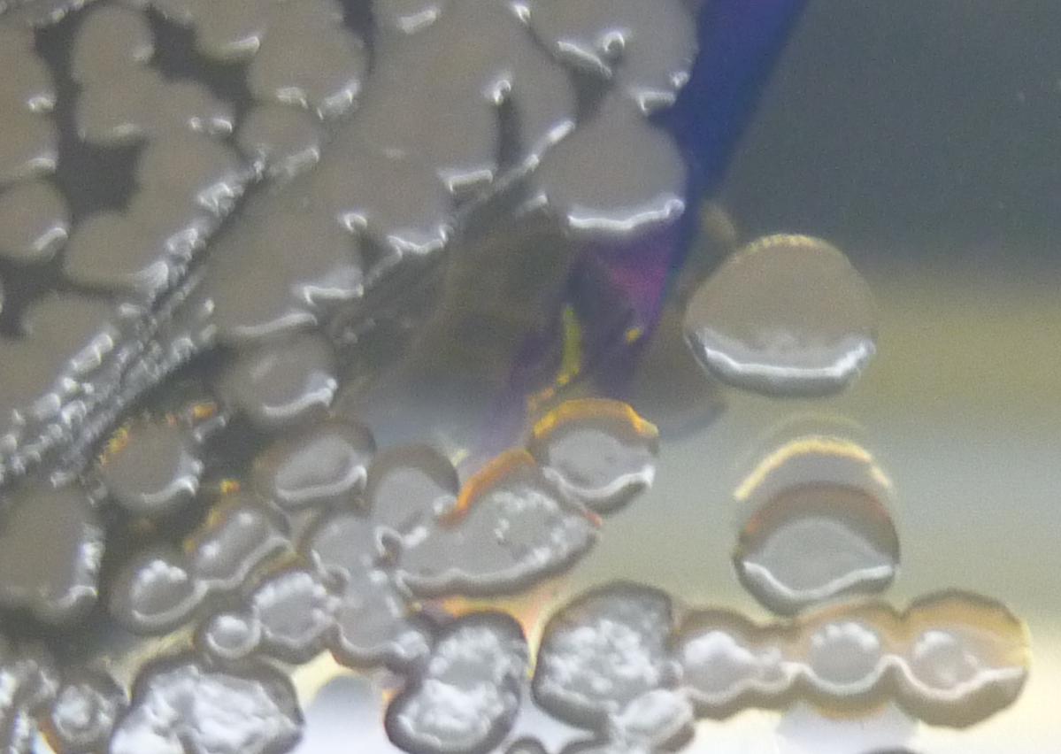
Constructs with chaplin E and H where present in Bacillus subtilis and Bacillus subtilis ΔTasA. 100uL of subtilin solution 0%, 0.1%, 1%, 10% and 100% where plated on LB agar plates and let dry for 10min. Each strain was spread out with an inoculation loop on each concentration. As a control Bacillus subtilis and Bacillus subtilis ΔTasA without chaplins were spread out on all concentrations as well. The next morning the plates where visually analyzed. No difference between different subtilin concentrations were visible, furthermore, there were no differences between the strains with chaplins and the ones without. Adding a drop of water on the colonies did not show any difference in repellence between any of the samples. The only recognizable difference was the colonies morphologies between strains with or without TasA. The ΔTasA colonies where smaller and shinier since TasA is involved in biofilm formation.
Geeske
Continued with the samples Jorrit took after induction. He stored the supernatant and pellet separately at -20ºC. I proceded with TCA (supernatant) and TFA (of supernatant after TCA and of lysed and speedvac’ed pellet samples) treatments and made samples to analyse with SDS-PAGE. Gels showed inconclusive results.
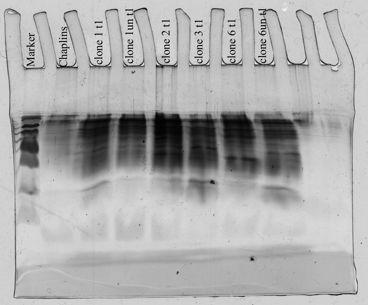 all samples are the supernatant samples
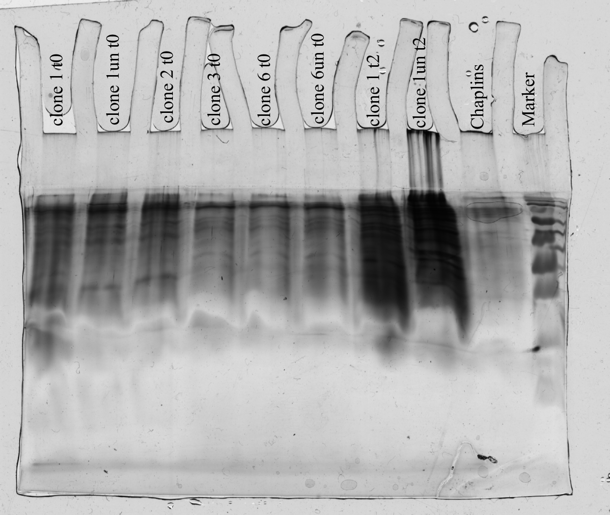 all samples are the supernatant samples
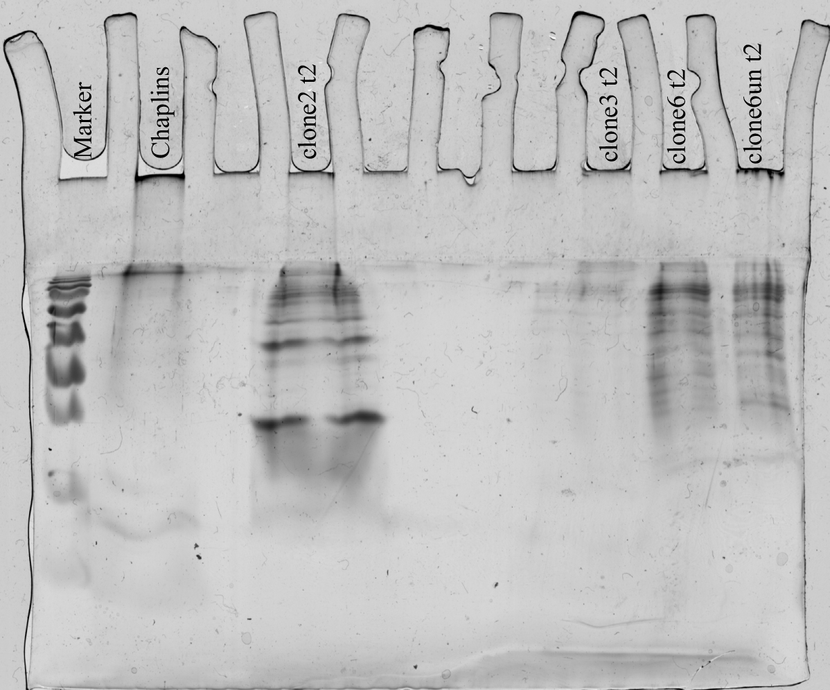
gel did not work as planned. samples leaked under other wells. all samples are the supernatant samples
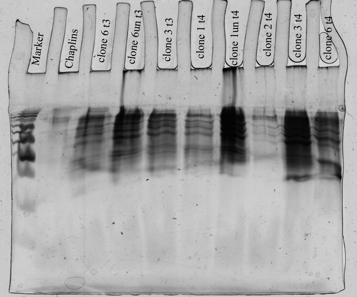 all samples are the supernatant samples
We need better controls like empty pNZ8901 and a GFP-control because it seems like the inducing-system is leaky. When chaplins assemble they stay in the stacking gel during SDS-PAGE (Data from pellet-samples not shown).
Modellers
Trying to understand the 3D biofilm model of the van Loosdrecht group and search for more information Joël, Laura, Djoke
 "
"

