Team:Cambridge/Notebook/Week8
From 2010.igem.org
(→Monday) |
(Replacing page with '{{:Team:Cambridge/Templates/HybridBook|num=8}}') |
||
| (One intermediate revision not shown) | |||
| Line 1: | Line 1: | ||
| - | {{:Team:Cambridge/Templates/ | + | {{:Team:Cambridge/Templates/HybridBook|num=8}} |
| - | + | ||
| - | + | ||
| - | + | ||
| - | + | ||
| - | + | ||
| - | + | ||
| - | + | ||
| - | + | ||
| - | + | ||
| - | + | ||
| - | + | ||
| - | + | ||
| - | + | ||
| - | + | ||
| - | + | ||
| - | + | ||
| - | + | ||
| - | + | ||
| - | + | ||
| - | + | ||
| - | + | ||
| - | + | ||
Latest revision as of 17:36, 2 September 2010

Viewing project diary
View protocols and data
|
Week 8: Monday 30th August - Sunday 5th September
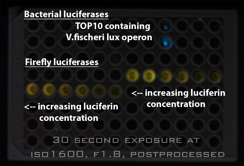
MondayAnja repeated lux operon extraction with pJS-555 instead of pSK-555 in the hope of getting better results. TuesdaySadly no light from the cells transformed with the extracted lux operon. WednesdayTheo attempted to test induction of pBAD in the BMG Labtech plate reader ThursdayWe came in in the morning and viewed the results from the plate reader experiment that had been running overnight. The bacterial lux results were disappointing. Our positive control was emitting (blue) light. (See photo on the right), but the other cells which had been incubated with arabinose in the hope of inducing them had not emitted light. But the most interesting results came from the cells expressing firefly luciferase ([http://partsregistry.org/Part:BBa_I712019 BBa_I712019]) under a consitutive promoter / RBS ([http://partsregistry.org/wiki/index.php?title=Part:BBa_J13002 BBa_J13002]. These showed low light output for 13 hours and then a rapid increase in luminescence, to a level which was sustained until the evening. We left the cells in the reader overnight to continue reading. FridayWe got results from sequencing of what we hoped to be our first Biobrick. It was not what we had hoped for, which was dissapointing. Additionally, Theo miniprepped a lux operon assembled using Gibson techniques and found it was the wrong size, after restriction enzyme cutting and running on a gel. But on a positive note synthesis from DNA 2.0 arrived, and preliminary results using CHBT combined with D-cysteine showed significant light output implying that functional LRE should allow recycling of oxyluciferin. SaturdaySundayTheo made a tool to allow export of BioBricks with annotations. |
Week 8: Monday 30th August - Sunday 5th September
MondayResult (from Expt. 68): No growth overnight, 2 growths out of 3 plates (+1 fungus) after weekend of growing. No glow. 69. Expt: Growing up of cell cultures of above experimental results & of results on p49 (Will)Strains G1, j1 and G28wh were grown up in 5ml LB + 2µl Chloramphenicol, and on plates containing LB+Cm. Left to grow at 30°C for 24h. Results: Strains grew but did not glow after 48h. 70. Expt: Miniprep pSB1C3 from colonies (Anja)Suspended a 'streak' of bacteria transformed with BBa_J04450 (registry part in pSB1C3) in 250µl buffer P1. Followed 'Bench protocol: Qiagen Spin Miniprep Kit Using a Microcentrifuge'. Nanodrop 12.8ng/µl 71. Expt: Gibson assembly of pBAD, luxCD,AB,EG and pSB1C3 (Anja)PCR:
BBa_J04450: once purified (E), once colony PCR (EC) Followed protocol on p38 for PCR mixtures and programs. Gel electrophoresisSelf-cast 1% agarose gel loaded as follows:
Run at 120V. No bands were visible. Not even in the marker lane. Since a gel was that had been sitting on the bench for a few days and SYBR Safe Dye is light sensitive it is assumed that the dye had become non-functional. A 1% Agarose gel was loaded as follows:
These were all mixed and spun down prior to gel loading. Bands of correct sizes were observed in all cases. Gel ExtractionDNA was extracted from the gel following the "Bench protocol: MinElute Gel Extraction Microcentrifuge protocol". Tuesday72. Expt: Continued Gibson assembly of pBAD, luxCD,AB,EG and pSB1C3 (Anja)Fresh Gibson 1.33x Master Mix was prepared following the protocol on p40. A Gibson Assembly reaction was prepared according to the protocol on p40, but taking twice the volume of the Master Mix (ie 30µl) and each fragment (ie 2µl). The reaction was incubated for 1h at 50°C. TransformationTOP10 cells, red strain and black strain were transformed with 10µl of Gibson reaction following protocol on p13+14. Plated 150µl on LB agar plates with Chl. (Since we were short of LB+Agar+Chl plates, two old LB agar + 5µg/ml Chl plates from PJ were used). Incubated at 30°C overnight. 73. Expt: Prepare DNA sequencing material for BioScience (Theo and Anja)Extracted plasmids from TOP10 cells were transformed with pBAD, luxCD,AB,EG in pSB1C3 following the 'Qiaprep Spin Miniprep Kit Using a Microcentrifuge' protocol. Experiment was cancelled (to be repeated on 01/09/10) Wednesday74. Expt: Prepare DNA sequencing material for BioScience (Anja)Extracted plasmids from TOP10 cells were transformed with pBAD, luxCD,AB,EG in pSB1C3 following the 'Qiaprep Spin Miniprep Kit Using a Microcentrifuge' protocol. Nanodrop measurements: 88.7ng/µl Primers are at 100µM = 100pmol/µl --> 30x dilution --> 3.2pmol/µl BioScience requests DNA concentrations of 100ng/µl for plasmids (Volume: 6µl) and 3.2pmol/µl (Volume: 10µl) for primers.
75. Expt: Repeat of PCR of pBAD+luxCDABEG using modified reaction conditions (Will)Added on ice:
notation as on p38
In addition EG was run in touchdown PCR.
PCR products were loaded into 1g/100ml agarose gel with 20µl/100ml SYBR SAFE strain. Per well 25µl product + 5µl Gel loading dye Wells: Hyperladder I, A, B, C, D, E, Dtouchdown, Hyperladder I 76. Night of 1 September - Plate ReaderIn two parts: (A) Arabinose induction of G28 (lux operon under pBAD) Rows A and B: all LB+Chl (100µl)
Incubated at 30°C Plate reader protocol:
Layout:
Thursday77. Expt: PCR of pBAD, luxCD,AB,EG and pSB1C3 (Will and Anja)PCR mixtures were made up according to p38. Notation as follows:
A,B and C were run at the following conditions:
D was run at:
Dp, E, Ec & Ep were run at:
Gel ElectrophoresisIn self-cast 1% agarose gels (with 30µl SYBR safe dye per gel) ran: 25µl PCR reaction + 5µl 6x orange LD (mixed prior to loading). Ladder: Hyperladder I A,B,C,E,Dp were cut out for gel extraction. To improve the yield of fragment E, bands from both gels were cut out and pooled. 78. Expt: Firefly Luciferase/Luciferin overnight plate reader experiments (+CHBT test) (Will)Plate reader: OD, 100ml, luminescence gain 3200, read length 10s, reads every 10 minutes, overnight (from 1st to 2nd Sept) Plate layout (X21 to 40):
'*' = 50µl broth + 50µl PBS All have 50µl cell in broth + 50µl substrate solution.
For 200µl:
Luciferin, sodium salt, molar mass = 302g/mol Stock at 2mg/µl = 0.002g/ml 0.002g/302g/mol = 6.62x10^-6 mol/ml or 6.62x10^-3 Molar solution (6.62mM) We want 0.4mM so 6.62/0.4 = 16.5x dilution ResultsCHBT: no light 79. Expt: Transforming reporters (Theo)Theo transformed TOP10cc with two translational units:
He plated out and made overnight cultures. 80. Expt: Gel extraction of pBAD, luxCD,AB,EG and pSB1C3 (continued from p65) (Will)Bands were cut out of the gels and DNA was extracted following the 'Bench protocol: MinElute Gel Extraction Microcentrifuge Protocol'. The extracted DNA was stored at -20°C overnight. 81. Testing colonies from transformation on 31.08.10 for presence of Gibson assembled plasmid (Anja)Colonies had grown on all plates from the transformation on 31.01.10, however, only after 3 days of incubating at 30°C. Are these colonies just a result of degraded chloramphenicol or are they containing the desired plasmid? A 10mM dNTP mix was prepared: 10µl 100mM dATP, dGTP, dTTP & dCTP + 60µl nuclease-free H20. Colony PCRMake up a master mix for 10 PCR reactions
Aimed to amplify luxEG region, used according to primers (see p38). Also prepared 10µM solutions of the two primers (using these rather than the 100µM stocks to reduce danger of contamination). The Master Mix was aliquated in 50µls, to which bacteria from one colony each were added (dip pipette tip 1st into colony then aliquated PCR mix, suck up PCR mix in pipette tip, pipette up and down a few times). PCR program (saved as BIOTAQ COL PCR) on G-storm:
Gel Electrophoresis1% Agarose E-gel was used. Loading mixtures contained 3µl 6x Orange LD, 7µl nuclease-free H20, 10µl PCR reaction mix There were lots of primer dimers => BEN WAS RIGHT! There is no band in any of the colonies of a size of ~1.8kb (corresponding to luxEG) => colonies do NOT contain desired plasmid :-( Friday82. Expt: Gibson assembly and transformation of pBAD, luxCD,AB,EG in pSB1C3 (continued from gel extraction on p.67) (Anja)Gibson assembly reaction
Incubated for 1h at 50°C TransformationTransformed 3x50µl TOP10 with 10µl Gibson reaction each. Following the protocol on p13+14. Plated 150µl on LB agar plates with Cm and incubated overnight at 37°C. Results: On the 04.09.10 two of three transformations were observed to yield colonies (ie colonies on two of three plates), most of plates which (not all!) were pink. ==> Need to identify what plasmid they contain. 83. Expt: Digestions of rbs Luc, pBAD Slock Lux (G28), rbs+EYFP, rbs+ECFP (Theo)Theo miniprepped these:
Digestion:
Fast digested for 30 mins Gel run with Hyperladder I. Interpretation:
Saturday84. Expt: Recovering P. phosphoreum from old cultures for making glycerol stocks (Peter)Grew up overnight culture from the old Falcon 4 in the morning the culture was turbid but not glowing on photos. Streaked out two of the old phosphoreum lawns to achieve single, glowing colonies.
Sunday85. Expt: Glycerol stocks of TOP10 with tetR repressed rbs+luc+pSB1C3 (Peter)Liquid cultures had grown in 37°C revolving incubator for two days. Broth appeared overgrown and sedimenting => resuspended sediment and streaked out onto Chl plates (37°C), placed liquid cultures into fridge. 6/9/10 put 9 single colonies in plate reader with some D-luciferin, after 6 hours ODs were up to ~1.5 but no glowing was detectable. |
 "
"

