Team:SDU-Denmark/project-p
From 2010.igem.org
(Difference between revisions)
(→K343007) |
(→K343007) |
||
| Line 26: | Line 26: | ||
<br> | <br> | ||
'''3. Videomicroscopy and computer analysis:'''<br> | '''3. Videomicroscopy and computer analysis:'''<br> | ||
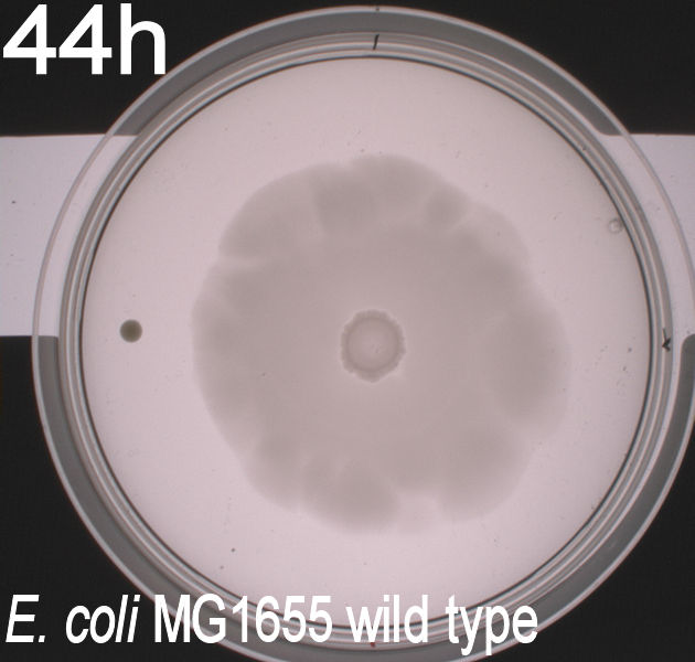
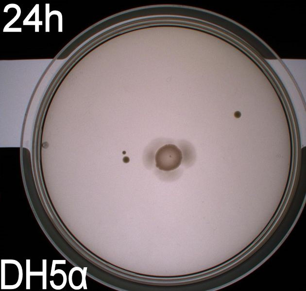
| - | To get an exact idea of what is happening to the bacteria when expressing the photosensor we decided on doing video microscopy of motile bacteria. | + | To get an exact idea of what is happening to the bacteria when expressing the photosensor we decided on doing video microscopy of motile bacteria. The bacterial cultures were prepared acording to protocol (insert link). Wildtype MG1655 and DH5alpha was used as controls. |
The actual microscopy was done on a (insert name) microscope with an optical magnification of 1000x. <br> | The actual microscopy was done on a (insert name) microscope with an optical magnification of 1000x. <br> | ||
To be able to observe an effect in the modified bacteria we had to expose the cultures to light, which was done through the microscope's integrated light source, which could switch between blue, green and ambient light. To ensure that the blue light was at the correct wavelength the same optical filter that was used for the plate experiments was inserted between the lightsource and the microscopy slide. Since microscopy is impossible without light, we had to use red light as a replacement for darkness, the source of the red light was ambient light from the microscope's light source filtered through a red optical filter (about 580nm). This should not have any effect on the phototactic bacteria according to Jung et. al (insert reference!). The room where the experiment was carried out was kept as dark as possible, as to eliminate the chance of the phototactic bacteria being unwantedly exposed to light. The actual experiment was carried out in the following manner:<br><br> | To be able to observe an effect in the modified bacteria we had to expose the cultures to light, which was done through the microscope's integrated light source, which could switch between blue, green and ambient light. To ensure that the blue light was at the correct wavelength the same optical filter that was used for the plate experiments was inserted between the lightsource and the microscopy slide. Since microscopy is impossible without light, we had to use red light as a replacement for darkness, the source of the red light was ambient light from the microscope's light source filtered through a red optical filter (about 580nm). This should not have any effect on the phototactic bacteria according to Jung et. al (insert reference!). The room where the experiment was carried out was kept as dark as possible, as to eliminate the chance of the phototactic bacteria being unwantedly exposed to light. The actual experiment was carried out in the following manner:<br><br> | ||
| Line 40: | Line 40: | ||
<object width="320"><param name="movie" value="http://www.youtube.com/v/HEKbYjyWNJc?hl=de&fs=1"></param><param name="allowFullScreen" value="true"></param><param name="allowscriptaccess" value="always"></param><embed src="http://www.youtube.com/v/HEKbYjyWNJc?hl=de&fs=1" type="application/x-shockwave-flash" allowscriptaccess="always" allowfullscreen="true" width="300"></embed></object> <object width="320"><param name="movie" value="http://www.youtube.com/v/bdg36HRHGRc?hl=de&fs=1"></param><param name="allowFullScreen" value="true"></param><param name="allowscriptaccess" value="always"></param><embed src="http://www.youtube.com/v/bdg36HRHGRc?hl=de&fs=1" type="application/x-shockwave-flash" allowscriptaccess="always" allowfullscreen="true" width="320" ></embed></object> </html><br><br> | <object width="320"><param name="movie" value="http://www.youtube.com/v/HEKbYjyWNJc?hl=de&fs=1"></param><param name="allowFullScreen" value="true"></param><param name="allowscriptaccess" value="always"></param><embed src="http://www.youtube.com/v/HEKbYjyWNJc?hl=de&fs=1" type="application/x-shockwave-flash" allowscriptaccess="always" allowfullscreen="true" width="300"></embed></object> <object width="320"><param name="movie" value="http://www.youtube.com/v/bdg36HRHGRc?hl=de&fs=1"></param><param name="allowFullScreen" value="true"></param><param name="allowscriptaccess" value="always"></param><embed src="http://www.youtube.com/v/bdg36HRHGRc?hl=de&fs=1" type="application/x-shockwave-flash" allowscriptaccess="always" allowfullscreen="true" width="320" ></embed></object> </html><br><br> | ||
When played at real time speed these videos seem slow and not showing any tendency, though when played at 4 times the normal speed, the photosensor will seem to show slightly increased motility when exposed to blue light. Even though the bacteria displayed disappointing motility, we went on and did computer analysis on the videos by the help of the open source software [http://db.cse.ohio-state.edu/CellTrack/ "CellTrack"]. The paths that were mapped showed a longer traveling distance for the bacteria expressing the photosensor compared to the rest of the samples when exposed to blue light. This might indicate a decreased tumbling frequency, but the low motility of the bacteria along with the inadequacy of the cell track software, the results of this experiments was rendered unreliable and a new protocol had to be devised.<br><br> | When played at real time speed these videos seem slow and not showing any tendency, though when played at 4 times the normal speed, the photosensor will seem to show slightly increased motility when exposed to blue light. Even though the bacteria displayed disappointing motility, we went on and did computer analysis on the videos by the help of the open source software [http://db.cse.ohio-state.edu/CellTrack/ "CellTrack"]. The paths that were mapped showed a longer traveling distance for the bacteria expressing the photosensor compared to the rest of the samples when exposed to blue light. This might indicate a decreased tumbling frequency, but the low motility of the bacteria along with the inadequacy of the cell track software, the results of this experiments was rendered unreliable and a new protocol had to be devised.<br><br> | ||
| - | '''4. Computerized analysis of the bacterial motility with the | + | '''4. Computerized analysis of the bacterial motility with the THOR prototype by [http://unisensor.dk/ Unisensor A/S] and the Unify software:'''<br> |
| - | For this experiment we changed our protocol for cultivating swimming bacteria | + | For this experiment we changed our protocol for cultivating swimming bacteria in order to optimize their mobility (insert link til protocol)<br> |
| - | + | The machine and software used for the analysis are prototypes and still under heavy development. Because of trade secrets it is impossible for us to explain how the machine works or give a detailed explanation of it's mechanism until the THOR has reached production status. Simplistically said it is a very advanced video microscope, with which it is possible to analyse liquids and the particles inside in both 2D and 3D, while also tracking them over time. | |
| - | The machine and software used for the analysis are prototypes and still under heavy development. Because of trade secrets it is impossible for us to explain how the machine works or give a detailed explanation of it's mechanism until the | + | 2.5 ul of the bacterial dilution were placed on the center of the microscopy slide and covered by a coverslide. Afterwards the sample was sealed with mineral oil as to prevent a flow in the liquid. The data collection was done exactly as described in the previously experiment, with the exeption that instead of optical filters we used diodes that emitted light at the wanted wavelengths (500nm and 600nm respectively). Moreover we also recorded the bacteria's behaviour when exposed to a blue light gradient and varying intensities of light measured in milliampere. All the measurements were carried out on the modified phototactic bacteria and the wildtype MG1655.<br> |
| - | 2.5 ul of the bacterial dilution were placed on the center of the microscopy slide and covered by a coverslide. Afterwards the sample was sealed with mineral oil as to prevent a flow in the liquid. The data collection was done exactly as described | + | |
The results look like these:<br> | The results look like these:<br> | ||
<br><html> | <br><html> | ||
<object width="320"><param name="movie" value="http://www.youtube.com/v/IlPRLSEG864?hl=de&fs=1"></param><param name="allowFullScreen" value="true"></param><param name="allowscriptaccess" value="always"></param><embed src="http://www.youtube.com/v/IlPRLSEG864?hl=de&fs=1" type="application/x-shockwave-flash" allowscriptaccess="always" allowfullscreen="true" width="320"></embed></object> <object width="320"><param name="movie" value="http://www.youtube.com/v/HC6DRAWcHys?hl=de&fs=1"></param><param name="allowFullScreen" value="true"></param><param name="allowscriptaccess" value="always"></param><embed src="http://www.youtube.com/v/HC6DRAWcHys?hl=de&fs=1" type="application/x-shockwave-flash" allowscriptaccess="always" allowfullscreen="true" width="320"></embed></object><br> | <object width="320"><param name="movie" value="http://www.youtube.com/v/IlPRLSEG864?hl=de&fs=1"></param><param name="allowFullScreen" value="true"></param><param name="allowscriptaccess" value="always"></param><embed src="http://www.youtube.com/v/IlPRLSEG864?hl=de&fs=1" type="application/x-shockwave-flash" allowscriptaccess="always" allowfullscreen="true" width="320"></embed></object> <object width="320"><param name="movie" value="http://www.youtube.com/v/HC6DRAWcHys?hl=de&fs=1"></param><param name="allowFullScreen" value="true"></param><param name="allowscriptaccess" value="always"></param><embed src="http://www.youtube.com/v/HC6DRAWcHys?hl=de&fs=1" type="application/x-shockwave-flash" allowscriptaccess="always" allowfullscreen="true" width="320"></embed></object><br> | ||
<br></html> | <br></html> | ||
| - | The bacteria that are not moving are assumed to be sticking to the glass or coverslip. | + | The bacteria that are not moving in the video are assumed to be sticking to the glass or coverslip. |
<br> | <br> | ||
Revision as of 14:47, 24 October 2010
 "
"
