Team:SDU-Denmark/project-p
From 2010.igem.org
(Difference between revisions)
(→K343007) |
(→K343007) |
||
| Line 26: | Line 26: | ||
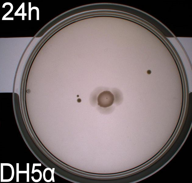
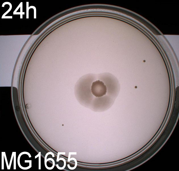
To get an exact idea of what is happening to the bacteria when adding the photosensor we decided on doing video microscopy of motile bacteria. The bacterial cultures for this analysis were incubated at 37° C, 180 RPM overnight, afterwards the optical density at 550nm was measured and the cultures were diluted to an OD550 of 0,02. They were then again incubated at 37 degrees and 180 RPM until their optical density reached 0,5. At this point we added retinal to the culture at a concentration of 5 uM and incubated the cells in the same conditions for another two hours. On top of phototactic bacteria we cultivated MG1655 (wildtype) and DH5alpha (non-motile) as controls. These were grown until an OD550 of 0,5 with the same methods as described for the modified culture, at this point these strains were ready for microscopy. <br> | To get an exact idea of what is happening to the bacteria when adding the photosensor we decided on doing video microscopy of motile bacteria. The bacterial cultures for this analysis were incubated at 37° C, 180 RPM overnight, afterwards the optical density at 550nm was measured and the cultures were diluted to an OD550 of 0,02. They were then again incubated at 37 degrees and 180 RPM until their optical density reached 0,5. At this point we added retinal to the culture at a concentration of 5 uM and incubated the cells in the same conditions for another two hours. On top of phototactic bacteria we cultivated MG1655 (wildtype) and DH5alpha (non-motile) as controls. These were grown until an OD550 of 0,5 with the same methods as described for the modified culture, at this point these strains were ready for microscopy. <br> | ||
The actual microscopy was done on a (insert name) microscope with an optical magnification of 1000x. <br> | The actual microscopy was done on a (insert name) microscope with an optical magnification of 1000x. <br> | ||
| - | To be able to observe an effect in the modified bacteria we had to expose the cultures to light, which was done through the microscope's integrated light source, which could switch between blue, green and ambient light. To further make sure that the blue light was at the correct wavelength the same optical filter that was used for the plate experiments was inserted between the lightsource and the microscopy slide. Since microscopy is impossible without light, we had to use red light as a replacement for darkness, the source of the red light was ambient light from the microscope's light source filtered through a red optical filter (about 580nm). This should not have any effect on the phototactic bacteria according to Jung et. al (insert reference!). | + | To be able to observe an effect in the modified bacteria we had to expose the cultures to light, which was done through the microscope's integrated light source, which could switch between blue, green and ambient light. To further make sure that the blue light was at the correct wavelength the same optical filter that was used for the plate experiments was inserted between the lightsource and the microscopy slide. Since microscopy is impossible without light, we had to use red light as a replacement for darkness, the source of the red light was ambient light from the microscope's light source filtered through a red optical filter (about 580nm). This should not have any effect on the phototactic bacteria according to Jung et. al (insert reference!). The microscopic slides were prepared by adding 5 ul of bacterial culture in the center of a glass microscopy slide, dimensions 7,5cm x 1,5cm, at last a cover slip was used to cover the culture. This was then sealed with ordinary nail polish to eliminate any flow in the system, which could be mistaken for bacterial motility. The room where the experiment was carried out was kept as dark as possible, as to eliminate the chance of the phototactic bacteria being unwantedly exposed to light. The actual experiment was carried out in the following manner:<br><br> |
| + | |||
| + | 1. Expose bacteria to red light for 1 minute, while recording video. | ||
| + | 2. Move to another spot on the slide, repeat 1. (repeated until 5 videos were recorded) | ||
| + | 3. Expose bacteria to blue light for 1 minute, while recording video. | ||
| + | 4. Wait for 30 seconds, for the bacteria to reset themselves. | ||
| + | 5. Move the focus to another spot on the slide, repeat 3. (x5)<br><br> | ||
| + | |||
| + | This was done with all three strains of bacteria.<br> | ||
| + | An example of the results: <br> | ||
| + | <object width="425" height="344"><param name="movie" value="http://www.youtube.com/v/HEKbYjyWNJc?hl=de&fs=1"></param><param name="allowFullScreen" value="true"></param><param name="allowscriptaccess" value="always"></param><embed src="http://www.youtube.com/v/HEKbYjyWNJc?hl=de&fs=1" type="application/x-shockwave-flash" allowscriptaccess="always" allowfullscreen="true" width="425" height="344"></embed></object> | ||
==== K343004 ==== | ==== K343004 ==== | ||
Revision as of 01:44, 24 October 2010
 "
"


