 "
"
Team:INSA-Lyon/Project/Stage1/Results
From 2010.igem.org
m |
|||
| Line 37: | Line 37: | ||
<h2>Results</h2> | <h2>Results</h2> | ||
<br><br> | <br><br> | ||
| - | <h3 style="color:purple;"><b>The | + | <h3 style="color:purple;"><b>The pILI1 plasmid</b></h3><br/> |
| - | <p>The transformed bacteria had been spread on LB plate with 7% of glucose and RedNile. | + | <p>The transformed bacteria had been spread on LB agar plate, with or without 7% of glucose and RedNile dye. As you can see on the photos below, the bacteria appear pink colored directly on plates supplemented with glucose. However, the control without glucose doesn't reveal a pink coloration.</p><br/> |
<div style="text-align:center;"> | <div style="text-align:center;"> | ||
<img src="http://lh4.ggpht.com/_Uc3bmii-yi0/TMf__pnKyLI/AAAAAAAAAl4/KBXck8LZerk/P1050468%20%282%29.JPG" /> | <img src="http://lh4.ggpht.com/_Uc3bmii-yi0/TMf__pnKyLI/AAAAAAAAAl4/KBXck8LZerk/P1050468%20%282%29.JPG" /> | ||
| Line 45: | Line 45: | ||
<img src="http://lh5.ggpht.com/_Uc3bmii-yi0/TMf-waXc1cI/AAAAAAAAAls/br-a_UaCThk/IMGP0983.JPG" /> <br/> | <img src="http://lh5.ggpht.com/_Uc3bmii-yi0/TMf-waXc1cI/AAAAAAAAAls/br-a_UaCThk/IMGP0983.JPG" /> <br/> | ||
| - | <p style="font-size:0.9em; text-indent:0px; text-align:center;"><em> | + | <p style="font-size:0.9em; text-indent:0px; text-align:center;"><em>Plates with pILI1 LB with or without Glucose 7% and RedNile dye.</em><br/><br/><br/></p> |
</div><br/> | </div><br/> | ||
| - | <p>With a liquid culture on LB complemented with glucose (7%) and RedNile, we observed the fluorescence by microscopy.</p><br/> | + | <p>With a liquid culture on LB complemented with glucose (7%) and RedNile dye, we observed the fluorescence by microscopy.</p><br/> |
<div style="text-align:center;"> | <div style="text-align:center;"> | ||
Revision as of 14:44, 27 October 2010
Results
The pILI1 plasmid
The transformed bacteria had been spread on LB agar plate, with or without 7% of glucose and RedNile dye. As you can see on the photos below, the bacteria appear pink colored directly on plates supplemented with glucose. However, the control without glucose doesn't reveal a pink coloration.
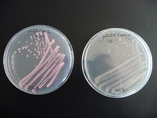

Plates with pILI1 LB with or without Glucose 7% and RedNile dye.
With a liquid culture on LB complemented with glucose (7%) and RedNile dye, we observed the fluorescence by microscopy.
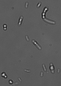
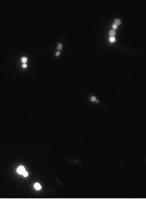
Observation of fluorescent granule by microscopy
The separated genes
We extracted the gene phaC from the plasmid of synthesis of Mr Gene. The gene had been successfully transferred in the pSBC3 plasmid. We send the product to the registry: BBa_K342001