Team:ETHZ Basel/Biology/Molecular Mechanism
From 2010.igem.org
(→Chemotactic network: Che proteins) |
(→Chemotactic network: Che proteins) |
||
| Line 11: | Line 11: | ||
This network consists of membrane (methyl accepting chemotaxis proteins: MCPs) and intracellular proteins (Che). MCPs sense the change in input concentration and communicate the information to the proteins CheW and CheA, located inside the cell. The autophosphorylation of CheA mediated by the MCPs is the key step in tumbling induction in response to increased repellent or decreased attractant concentration. The methylation state of the MCPs is influenced by the methyltransferase '''CheR''' and the demethylase '''CheB'''. CheA phosphorylates '''CheY''' which then diffuses through the cytoplasm to the flagellar motor protein FliM where it induces tumbling. The phosphatase '''CheZ''' regulates the signal termination through dephosphorylation of CheYp [[Team:ETHZ_Basel/Biology/Molecular_Mechanism#References|[5]]]. | This network consists of membrane (methyl accepting chemotaxis proteins: MCPs) and intracellular proteins (Che). MCPs sense the change in input concentration and communicate the information to the proteins CheW and CheA, located inside the cell. The autophosphorylation of CheA mediated by the MCPs is the key step in tumbling induction in response to increased repellent or decreased attractant concentration. The methylation state of the MCPs is influenced by the methyltransferase '''CheR''' and the demethylase '''CheB'''. CheA phosphorylates '''CheY''' which then diffuses through the cytoplasm to the flagellar motor protein FliM where it induces tumbling. The phosphatase '''CheZ''' regulates the signal termination through dephosphorylation of CheYp [[Team:ETHZ_Basel/Biology/Molecular_Mechanism#References|[5]]]. | ||
| - | The [https://2010.igem.org/Team:ETHZ_Basel/Modeling/Movement | + | The [https://2010.igem.org/Team:ETHZ_Basel/Modeling/Movement movement model] developed by our team simulates the motility of E. coli, by probabilistically switching between the two states: directed movement and tumbling, as a function of the CheYp concentration received as input from the [https://2010.igem.org/Team:ETHZ_Basel/Modeling/Chemotaxis chemotaxis pathway model]. |
= Structure of the Che proteins = | = Structure of the Che proteins = | ||
Revision as of 11:57, 23 October 2010
Molecular Mechanism
Chemotactic network: Che proteins
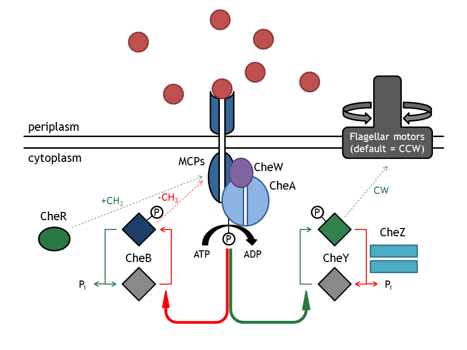
The chemotactic network is responsible for the directed movement of the bacterium, as a response to changes in the extracellular chemoattractant or chemorepellent concentration. The two types of movement that the bacterium can employ are tumbling (i.e. change in angle and no change in spatial coordinates), occurring when the bacterium doesn’t sense an increase in attractant concentration and directed movement ( i.e. change in position and slight change in angle), occurring when the attractant concentration is sensed as increasing. In E. coli, these two types of movements correspond to different rotation directions of its 4 flagellar motors (clockwise: tumbling; counter-clockwise: directed movement).
This network consists of membrane (methyl accepting chemotaxis proteins: MCPs) and intracellular proteins (Che). MCPs sense the change in input concentration and communicate the information to the proteins CheW and CheA, located inside the cell. The autophosphorylation of CheA mediated by the MCPs is the key step in tumbling induction in response to increased repellent or decreased attractant concentration. The methylation state of the MCPs is influenced by the methyltransferase CheR and the demethylase CheB. CheA phosphorylates CheY which then diffuses through the cytoplasm to the flagellar motor protein FliM where it induces tumbling. The phosphatase CheZ regulates the signal termination through dephosphorylation of CheYp [5].
The movement model developed by our team simulates the motility of E. coli, by probabilistically switching between the two states: directed movement and tumbling, as a function of the CheYp concentration received as input from the chemotaxis pathway model.
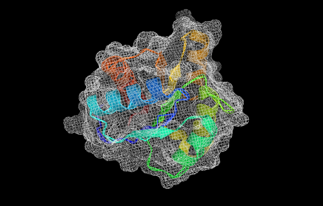
Structure of the Che proteins
CheY
CheR
Light activated system: PhyB-Pif3
In plants and some bacteria, members of the photoreceptor family of phytochromes regulate phototaxis, photosynthesis and production of protective pigments in response to light stimuli. The chromophore is encoded in the N-terminal domain of the protein [1]. There are two mayor types of phytochromes, type I (PhyA) and type II (PhyB and PhyC) [2, p.142]. Both exist in a biologically inactive form Pr absorbing red light and an active configuration Pfr which absorbs far-red light using a covalently attached tetraphyrrole chromophore for light absorption [5]. The interconversion between these two states takes only miliseconds [3]. While the Pr form is very stable (half live of about 100h), the Pfr form is faster degraded (type I half-life between 30min and 2h, type II half live around 8h) [2, p.142 ff].
Light-switchable gene systems utilize the phytochrome interacting factor Pif3, a basic helix-loop-helix protein, for light-induced gene expression. The N-Terminus of Pif3 selectively binds the Prf form of the phytochrome and rapidely dissociates in response to reconversion to the Pr state [3].
For the design of a light-activated system in Bacteria one has to consider that chromophores such as phycocyanobilin PBS are not naturally present; therefore, they either have to be added to the media or two genes for its biosynthesis starting from Haem have to be introduced [4]. Furthermore, the fact that phyotochromes can form sequesters in the Prf state in the timescale of seconds has to be examined [2].
Spatial localization in a bacterial cell: Anchor proteins
By spatial localization of one Che protein, the tumling frequency of a bacterial cell is influenced.
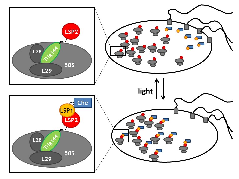
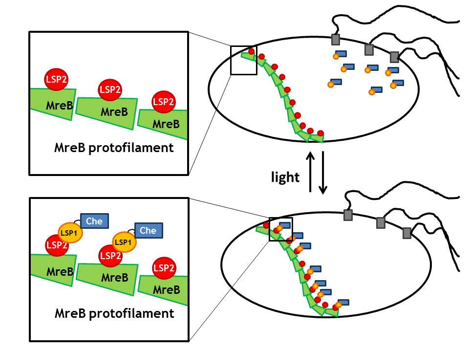
Such anchoring can either be achieved by fusion of the light sensitive protein to the tetracyclin repressor tetR, which binds to it's operator tetO present on a high copy vector in the cell [6]. Another possiblility is the fusion to the ribosome binding domain of the trigger factor trigA that binds to the large subunit of the ribosome [7]. The prokaryotic actin homologue MreB represents en alternative enabling the localization of the light sensitive protein to the cell wall [8].
Upon light signal, the light-sensitive proteins LSP's dimerize and therefore spatial localize the fused Che-protein affecting the activity of the Che downstream partners. Thus, these anchors enable induction and repression of running or tumbling.
References
[1] Sato, Hug, Tepperman and Quail: A light-switchable gene promoter system. Nature Biotechnology. 2002; 20.
[2] Kendrick and Kronenberg: Photomorphogenesis in plants. Kluwer academic publishers, Dordrecht, The Netherlands. 2nd edition, 1994.
[3] Keyes and Mills: Inducible systems see light. Trends in Biotechnology. 2003; 21:2.
[4] Levskaya, Chevalier, Tabor, Simpson, Lavery, Levy, Davidson, Scourast, Ellington, Marcotte and Voigt: Engineering Escherichia coli to see light. Nature Brief Communications. 2005, 438.
[5] M.J. Tindall, S.L. Porter, P.K. Maini, G. Gaglia, J.P. Armitage. Bulletin of Mathematical Biology (2008) 70: 1525–1569.
[6] Bertram and Hillen: The application of Tet repressor in prokaryotic gene regulation and expression. Microbial Biotechnology. 2008; 1:1.
[7] Hesterkamp, Deuerling and Bukau: The Amino-terminal 118 amino acids of Escherichia coli Trigger factor constitute a domain that is necessary and sufficient for binding to ribosomes. The Journal of Biologcial Chemistry. 1997; 272:35.
[8] Kruse, Bork-Jensen and Gerdes: The morphogenetic MreBCD proteins of Escherichia coli form an essential membrane-bound complex. Molecular Microbiology. 2005;55:1.
 "
"


